Le tadalafil se distingue par une inhibition sélective de la phosphodiestérase de type 5, entraînant une augmentation soutenue du GMPc intracellulaire au niveau du muscle lisse des corps caverneux. Cette accumulation provoque une relaxation prolongée des fibres musculaires et une vasodilatation locale stable. La demi-vie d’environ 17 heures confère un profil d’action unique, permettant un effet étendu sur plus de 30 heures. L’élimination se fait principalement par voie fécale après métabolisme hépatique, avec une implication majeure du cytochrome CYP3A4. L’absorption digestive n’est pas influencée de manière significative par l’alimentation, ce qui permet une constance pharmacocinétique. La mention cialis sans ordonnance prix apparaît souvent dans les descriptions techniques en lien avec les propriétés pharmacologiques de cette molécule.
Dierenfaculteitnr04aug2010:dierenfaculteit
280 Case report
Vlaams Diergeneeskundig Tijdschrift, 2010, 79
Estrogen-induced pancytopenia due to a Sertoli cell tumor in a cryptorchid Beauceron Oestrogeengeïnduceerde pancytopenie door een sertoliceltumor bij een1 H. De Bosschere, 2 C. Deprest
1 Medisch Labo Bruyland – Afdeling Diergeneeskunde, Meiweg 1, B-8500 Kortrijk, Belgium2 Dierenartsenpraktijk Deprest, Verlovestraat 4, B-8755 Ruiselede, Belgium
ABSTRACT The present report describes the clinical history, radiography, ultrasonography, hematology, bone marrow sample, and histology of a case of estrogen-induced pancytopenia due to neoplastic change of cryptorchid testes. The anemia gradually improved after castration, several blood transfusions and treatment with nandrolondecanoate.
Dit is een beschrijving van de voorgeschiedenis, radiografie, echografie, hematologie, beenmergpunctie en
histologie van een oestrogeengeïnduceerde pancytopenie ten gevolge van een tumorale ontaarding van cryptorchetestikels. Deze anemie verbeterde geleidelijk na castratie, verscheidene bloedtransfusies en nandrolondecanoaat.
trophy or perianal adenoma, or when it is produced by
Pancytopenia is a medical condition in which there
a testicular tumor or ovarian granulose cell tumor (Son-
is a reduction in the numbers of red and white blood cells
tas et al., 2009). The response of the bone marrow to
and of platelets due to damage to stem cells or to the
estrogens varies from one dog to another, because some
bone marrow microenvironment leading to bone mar-
dogs may have fatal bone marrow suppression, where-
row failure. The frequently used term ‘aplastic anemia’
as others have only mild to moderate damage (Crafts,
is confusing since all cell lines are affected, and not only
the red blood cells. Bone marrow damage may be caused
Clinical signs relate to thrombocytopenia (petechial
by infections, drugs, toxins, neoplasia, myelodysplasia,
hemorrhages, melena, etc.), neutropenia (pyrexia and
bone marrow necrosis, osteosclerosis, myelofibrosis or
sepsis) and anemia (lethargy and pallor).
immune-mediated mechanisms, or it may be idiopath-
The present case report describes the successful treat-
ic (Harvey, 1997; Tilley and Smith, 2004; Villiers and
ment of a paraneoplastic syndrome of estrogen-induced
Blackwood, 2005). In the acute form of the disease, the
pancytopenia due to a Sertoli cell tumor in cryp-
destruction of progenitor and dividing cells leads to leu-
copenia/neutropenia within 5 days and to thrombocy-topenia within 8-10 days. Anemia develops more
gradually due to the long red blood cell lifespan. Bonemarrow aspiration reveals a mix of necrotic lysing cells,
Case history
macrophages and stromal cells. Depending on the cause,the bone marrow may recover and be repopulated, usu-
A 9-year-old bilateral cryptorchid male Beauceron
ally within 3 weeks after the original marrow injury, or
was presented in poor general condition. The owner was
the disease may progress to the chronic form. In the
aware of the possible risks of cryptorchidy, but refused
chronic form, the stem cell damage is irreversible and
to castrate the dog. During the examination, the dog did
the red marrow is replaced by fat, leading to neutropenia,
not show any obvious signs of feminizing syndrome,
thrombocytopenia and moderate to severe anemia
such as gynecomastia or alopecia. However, based on
the anamnesis of approximately one year prior to the
Estrogen-induced pancytopenia is only observed in
time of presentation, the dog had then been making cop-
dogs and ferrets (Crafts, 1948; Kociba and Caputo,
1981). The dog is very susceptible to the myelotoxic ef-
anamnesis, however, there was no report of abnormal
fects of estrogen when estrogen is administered in fe-
sexual behavior. Clinical examination revealed high fever
male dogs for urinary incontinence, mismating, estrus
(40.5°C) and increased heart beat (150 bpm). The dog
induction or pseudopregnancy, when it is administered
had a lick granuloma on the hind leg and a large bite
in male dogs for the management of prostatic hyper-
wound in the neck. The dog was treated conservative-
Vlaams Diergeneeskundig Tijdschrift, 2010, 79
Table 1. Results of blood analysis of a 9-year-old bilateral cryptorchid Beauceron. Hematology References 17-beta-estradiol
ly with antibiotics and nonsteroidal anti-inflammatory
incision from the umbilicus to the pubis. Both neoplastic
drugs (NSAIDS). Nine days later there was only slight
testicles were double ligated with Vicryl 2/0, transect
improvement in the dog’s condition. The dog was ane-
and removed. The abdomen was closed using a standard
mic and showed syncope after jumping. Physical ex-
procedure. Both testicles were histopathologically ex-
amination showed a temperature of 39.3°C, a systolic
heart murmur grade 2/6 left apex due to the anemic sta-tus of the patient, and weak femoral pulses. Other ex-
Histopathology of the testes
aminations such as blood analysis, radiography and ul-trasonography were carried out.
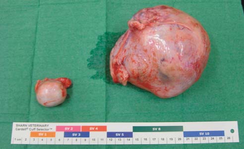
The size of the testes was respectively, 3.5 cm in di-
ameter and 11 cm by 7 cm (Figure 3). Hematoxilin eosin
Blood examination
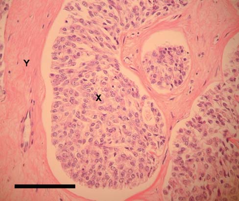
(HE) sections of the enlarged testis revealed a prolif-eration of Sertoli cells, spread over numerous tubuli, in-
The results of the hematology and follow-up analy-
termixed with a trabecular collagenous network. The
ses are summarized in Table 1. Initially, there was a mod-
neoplastic cells formed tubules of elongated cells in a
erate nonregenerative normocytic slightly hypochromic
parallel arrangement, with a large round to ovoid nu-
anemia, leucepenia/neutropenia and severe thrombo-
cleus and a large amount of vacuolated or dense
cytopenia. The 17-beta-estradiol concentration was
eosinophilic cytoplasm. The tumor consisted of well
markedly increased at 149.2 pg/ml (normal values 13-55 pg/ml). Radiography
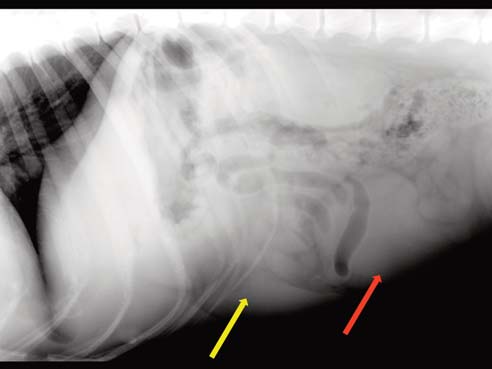
The x-ray of the thorax showed a normal heart with
a VHS (vertebral heart score) < 10.5 and a slightly di-minished lung vessel pattern. The x-ray of the abdomenshowed an enlarged spleen with a caudal well circum-scribed mass. The intestines were pushed dorsally andlaterally by this mass situated on the midventral aspectof the caudal abdomen (Figure 1).
Ultrasonography
On the ultrasonographic examination of the abdomen,
the mass noticed on the x-ray consisted of two mass-es, both showing echogenic zones. The larger of the two
Figure 1. The X-ray with the dog in right lateral recum-
masses was approximately 7 cm by 6 cm, while the oth-
bency shows an enlarged spleen (yellow arrow) and a well –circumscribed soft-tissue abdominal mass on the
er mass was smaller (Figure 2). These masses were sus-
caudal midventral abdomen (red arrow) approximately
pected to be neoplastic transformations of the cryptorchid
twice the size of the kidneys. The intestines seem to be
testes. Both testes were removed via a ventral midline
displaced dorsally and laterally of this mass.
Vlaams Diergeneeskundig Tijdschrift, 2010, 79
Figure 4. Neoplastic proliferation of Sertoli cells (X), spread over numerous tubuli, intermixed with a dense Figure 2. On ultrasonography, the mass observed on X- trabecular collagenous network (Y). The neoplastic cells ray appears to be well circumscribed with irregular hy- formed tubules lined by multiple layers of elongated cells poechoic zones (yellow arrow), suggestive of fluid. The in a parallel arrangement, with a large round to ovoid testicular parenchyma is mixed hypoechoic. nucleus and a large amount of eosinophilic cytoplasm. The cells are arranged perpendicularly to the basement membrane. HE staining - Bar = 100µm. Figure 3. The left testicle changed into the largest tumor and measured 11 cm by 7 cm, the right testicle tumor measured 3.5 cm in diameter.
formed tubules that were lined by multiple layers of neo-plastic Sertoli cells. These Sertoli cells were arranged
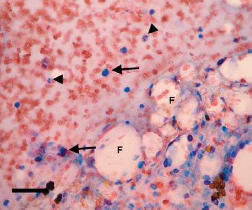
Figure 5. The bone marrow showed severe lack of ery-
perpendicularly to the basement membrane (Figure 4). throid, myeloid and megakaryocytic cell types. There
The contralateral smaller atrophic testis showed severe
were numerous mature erythrocytes, a moderate num- ber of neutrophils (arrowheads) and a moderate num-
fibrosis with a small number of tubular sections. ber of large mononuclear cells (arrows). Presence of fat
After surgery the anemia became worse (Table 1). (F). Modified Wright stain - Bar = 50 µm
Due to the nonregenerative nature of the anemia, a bonemarrow aspiration was carried out for evaluation. Treatment Bone marrow cytology
A diagnosis of estrogen-induced pancytopenia due
to a Sertoli cell tumor in a cryptorchid testis was made.
The bone marrow a showed severe lack of erythroid,
After castration, the dog received 4 consecutive blood
myeloid and megakaryocytic cell types. There were nu-
transfusions of 350 ml, each time at an interval of 4 to
merous mature erythrocytes, a low number of neutrophils
6 days. The 2nd and 4th blood transfusions were followed
with some band neutrophils, and a low number of large
by injections of Deca-Durabolin (200 mg IM - nan-
mononuclear cells (Figure 5). Small amounts of fat tis-
drolondecanoate). Over time, the anemia gradually
sue were also observed on the slides. These findings are
changed into a slightly regenerative macrocytic
consistent with estrogen-induced myelotoxicity.
hypochromic anemia, and thrombocytopenia. Fourmonths after the start of the treatment, the complete
Vlaams Diergeneeskundig Tijdschrift, 2010, 79
blood count was almost within reference levels (Table
infections, and stimulation of the remaining bone mar-
row (Hall, 1992). Untreated cases of estrogen inducedmyelotoxicity always have an unfavorable prognosis.
Death occurs from complications of hemorrhage and in-fection.
Sertoli cell tumors of the testes arise from the sup-
However, in chronic cases, the stem cell damage may
porting cells within the seminiferous tubuli. This is a
be irreversible and the red marrow is replaced by fat,
common neoplasia in dogs, especially in dogs with cryp-
leading to neutropenia, thrombocytopenia and moder-
torchid testicles. Almost 50% of all canine Sertoli cell
ate to severe anemia (Villiers and Blackwood, 2005).
tumors arise in cryptorchid testes and the incidence of
The use of lithium appeared to be successful in in-
Sertoli cell tumors is more than 20 times higher in cryp-
ducing regeneration of the bone marrow in dogs (Hall,
torchid than in scrotal testes (Hayes and Pendergrass,
1992). In humans, various drugs have been used suc-
1976). Approximately 20 to 30% of dogs with Sertoli
cessfully to treat aplastic anemia: corticosteroids, an-
cell tumor manifest signs of hyperestrogenism, char-
drogens, lithium carbonate and cyclophosphamide.
acterized by any combination of feminization, gy-
Bone marrow transplantation is also another possibil-
necomastia, atrophy of the contralateral testicle (as in
ity in humans (Kjeldsberg et al., 1989).
the present case), squamous metaplasia within the
In the present case, Deca-Durabolin (nandrolonde-
prostate gland (often with accompanying suppurative
canoate) was used. This drug is an anabolic steroid and
prostatitis), alopecia and bone marrow atrophy (as in the
is useful in treating certain types of anemia, such as
present case) (McEntee, 1990). It has not been proven
aplastic anemia and anemia due to chronic kidney fail-
that estrogen is solely responsible for all these mani-
ure. In certain cases where the bone marrow has
festations. Serum estrogen concentrations are not in-
stopped producing new erythrocytes, administration of
creased in some dogs with apparent hyperestrogenism
anabolic steroids will stimulate this system again and
associated with testicular Sertoli cell tumor (Grooten-
bring the numbers of these cells back to normal levels.
huis et al., 1990). However, in the present case the es-
They also are known to stimulate the production of white
trogen concentration was markedly increased. Estrogen-
blood cells and platelets, though to a lesser degree. In
induced production of myelopoiesis-inhibitory factor by
summary, the present dog showed a severe estrogen-
thymic stromal cells has been described (Grootenhuis
induced pancytopenia due to neoplastic transformation
et al., 1990; Farris and Benjamin, 1993). The bone mar-
of cryptorchid testes (paraneoplastic syndrome), which
row suppressive effects accompanying Sertoli cell tu-
gradually improved to almost within reference ranges
mors, also known as aplastic anemia, can be so severe
after castration, blood transfusions and administration
as to cause anemia, leukopenia and thrombocytopenia.
Aplastic anemia is characterized as a nonregenera-
tive anemia with normal erythrocytic indices, neu-
tropenia, and thrombocytopenia, and it suggests amultipotential stem cell disorder. Cytologic evaluation
We would like to thank the staff of Medisch Labo
of the bone marrow of the present case confirmed bone
Bruyland and Labo Pathologie Yperman for their as-
marrow aplasia. Neutropenia from decreased cellular
production usually has a minimal left shift (due to anincreased demand for neutrophils and/or depletion of
the bone marrow storage reserves of mature neu-trophils).
Crafts R. C. (1948). The effects of estrogens on the bone mar-
Differential diagnosis of nonregenerative anemia in-
row of adult female dogs. Blood 3, 276–285.
cludes: infections (canine parvovirus, Ehrlichia canis),
Farris G. M., Benjamin S. A. (1993). Inhibition of
drugs (estrogens, meloxicam, griseofulvin, chemother-
myelopoiesis by serum from dogs exposed to estrogen. American Journal of Veterinary Research 54, 1374-1379.
apy, phenylbutazone, trimethoprim/sulphonamine),
Grootenhuis A. J., van Sluijs F. J., Klaij I. A., Steenbergen
and idiopathic immune-mediated endogenous estrogen
J., Tillerman M. A., Bevers M. M. Dieleman S. J., de Jong
(Harvey, 1997; Villiers and Blackwood, 2005). Estro-
F. H. (1990). Inhibin, gonadotrophins and sex steroids in
gen toxicosis is a common cause of myelosuppression
dogs with Sertoli cell tumors. Journal of Endocrinology 127,
in the dog. This condition may result from a paraneo-
plastic syndrome (Sertoli cell tumor that produces es-
Hall E. J. (1992). Use of lithium for treatment of estrogen-
trogen), as in the present case, or from the administra-
induced bone marrow hypoplasia in a dog. Journal of theAmerican Veterinary Medical Association 200, 814-816.
The clinical outcome of reported cases of estrogen
Harvey J. W. (1997). The erythrocyte. In: Kaneko J. J., Har-
toxicity has either been death or a long recovery peri-
vey J. W., Bruss M. L. (eds.). Clinical Biochemistry of Do-
od (Sontas et al., 2009). Therapeutically, all the estro-
mestic Animals. 5th Ed., Academic Press, San Diego, pp. 166–167.
gen induced effects may disappear after removal of the
Hayes H. M., Pendergrass T., W. (1976). Canine testicular tu-
underlying cause of the myelotoxicity, i.e. castration in
mors: epidemiologic features of 410 dogs. International
the present case. Treatment also includes: correction of
Journal of Cancer 18, 482–487.
the anemia and thrombocytopenia, protections against
Kjeldsberg C., Beutler E., Bell C., Hougie C., Foucar K., Sav-
Vlaams Diergeneeskundig Tijdschrift, 2010, 79
age R. (1989). Aplastic, hypoplastic and miscellaneous types
Tilley L. P., Smith F. W. K. (2004). Estrogen toxicity. In: Tilley
of anemia. In: Practical Diagnosis of Hematologic Dis-
L. P., Smith F. W. K. (eds.). The 5-Minute Veterinary Con-orders. Revised edition, American Society of Clinical
sult: Canine and Feline. 3rd Ed., Philadelphia, Lippincott
Williams and Wilkins, pp. 430–431.
Kociba G. J., Caputo C. A. (1981). Aplastic anemia associ-
Villiers E., Blackwood L. (2005). Disorders of erythrocytes.
ated with estrus in pet ferrets. Journal of the American Vet-
In: BSAVA Manual of Canine and Feline Clinical Pathol-erinary Medical Association 178, 1293-1294. ogy. 2nd Ed., British Small Animal Veterinary Association,
McEntee K. (1990). Scrotum, spermatic cord and testis: pro-
liferative lesions. In: Reproductive Pathology of Domes-tic Animals. Academic Press Inc., San Diego, pp. 279–306.
Sontas H. B., Dokuzeylu B., Turna O., Ekici H. (2009). Es-
trogen-induced myelotoxicity in dogs: a review. Canadi-an Veterinary Journal 50, 1054 – 1058.
Domperidon zur Milchmengensteigerung bei stillenden Frauen Gudrun von der Ohe, Ärztin und IBCLC aus Hamburg Hintergründe Medikamente oder traditionelle „Hausmittelchen“ zur Milchmengensteigerung bei stillenden Frauen gibt es in allen Kulturen. Sie werden als Galaktogogen bezeichnet, sind in den meisten Fällen pharmakologisch wenig hilfreich, stehen aber immer mit einer
Prophylaxe- und Fachassistent/innen, Dentalhygieniker/innen ZMP / ZMF / DH Vortragsprogramm Dr. Alexander Dorsch Notfallmanagement in der Zahnarztpraxis Prof. Dr. Andrea Mombelli Die fortgeschrittene Parodontitis und die Rolle der ZMP/ZMF bzw. DH Prof. Dr. Andrea Mombelli Periimplantäre Infektionen und die Rolle der ZMP/ZMF bzw. DH Andrea Busch Prophylaxe bei Schwangeren: Wissensc
 Vlaams Diergeneeskundig Tijdschrift, 2010, 79
Table 1. Results of blood analysis of a 9-year-old bilateral cryptorchid Beauceron.
Vlaams Diergeneeskundig Tijdschrift, 2010, 79
Table 1. Results of blood analysis of a 9-year-old bilateral cryptorchid Beauceron. 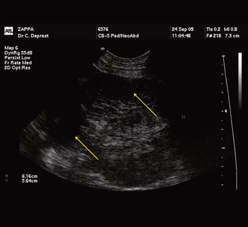
 Vlaams Diergeneeskundig Tijdschrift, 2010, 79
Figure 4. Neoplastic proliferation of Sertoli cells (X),
Vlaams Diergeneeskundig Tijdschrift, 2010, 79
Figure 4. Neoplastic proliferation of Sertoli cells (X),