Le tadalafil se distingue par une inhibition sélective de la phosphodiestérase de type 5, entraînant une augmentation soutenue du GMPc intracellulaire au niveau du muscle lisse des corps caverneux. Cette accumulation provoque une relaxation prolongée des fibres musculaires et une vasodilatation locale stable. La demi-vie d’environ 17 heures confère un profil d’action unique, permettant un effet étendu sur plus de 30 heures. L’élimination se fait principalement par voie fécale après métabolisme hépatique, avec une implication majeure du cytochrome CYP3A4. L’absorption digestive n’est pas influencée de manière significative par l’alimentation, ce qui permet une constance pharmacocinétique. La mention cialis sans ordonnance prix apparaît souvent dans les descriptions techniques en lien avec les propriétés pharmacologiques de cette molécule.
Combined intramedullary and external skeletal fixation of metatarsal and metacarpal fractures in 12 dogs and 19 cats
Combined Intramedullary and External Skeletal Fixation of Metatarsal and Metacarpal Fractures in 12 Dogs and 19 Cats
Noel Fitzpatrick1, DUniv MVB CertSAO CertVR, Jerry O. Riordan1, MVB CertSAS, Thomas J. Smith1, MAVetMB CertSAS, Jola H. Modlinska1, Russell Tucker2, DVM Diplomate ACVR, and Russell Yeadon1, MAVetMB CertSAS
1 Fitzpatrick Referrals,Halfway Lane, Eashing UK and 2College of Veterinary Medicine, Washington State University,Pullman, WA
Corresponding Author Objective: To report surgical technique, clinical experiences, and long-term out-
comes of combined intramedullary/external skeletal fixation of metatarsal (MT)
Halfway Lane, Eashing, Surrey, GU7 2QQ.
and metacarpal (MC) fractures in dogs and cats. Study Design: Case series. Animals: Dogs (n = 12); 19 cats. Methods: Clinical and radiographic records of animals managed by combined
intramedullary/external fixation of MT/MC fractures were reviewed. Signal-
ment, fracture configuration, complications, and subjective clinical findings were
recorded. Surgical technique involved retrograde intramedullary pin placement into fractured MT/MC bones, and transverse pin placement in the base of the MT/MCs or tarsal/carpal bones. Contoured pin ends were enshrouded dorsally in epoxy resin and implants maintained until fracture union. Postoperative clinical and radiographic reassessment was performed where possible. Results: Small breed dogs (n = 12) and 19 cats were operated. Fixator removal occurred in < 10 weeks in all cases. Complications included synostosis (n = 2), pin tract discharge (7), excessive postoperative swelling (8), skin abrasions from the frame (2), and paw distortion associated with frame impingement (2). Long-term radiography documented degenerative changes associated with MT-phalangeal or MC-phalangeal joints in 2 dogs; 7 cats, but changes were typically graded mild or moderate and affected only 1 or 2 joints. Conclusions: Combined intramedullary/external fixation of MT/MC fractures is viable, particularly juxta-articular fractures. Pin penetration of MT-phalangeal or MC-phalangeal joints may cause morbidity and requires further study.
Metatarsal (MT) and metacarpal (MC) injuries reportedly
rograde intramedullary pinning techniques7,12 whereby the
account for 8.1% and 3.3% of fractures in dogs and cats,
pin is driven distally through the head of the MT or MC
respectively,1 and are typically traumatic in origin.2 Mul-
bone, before being driven into the proximal fracture seg-
tiple MT or MC bones may be fractured, with fracture of
ment. Normograde insertion through a separately created
all 4 MT or MC bones in 41–56% of dogs.2,3 The mid-
“slot” in the dorsal aspect of the head of the MT or MC
to-distal MC or proximal MT regions are most frequently
bone may avoid the distal articular surfaces.9,13 This may
be technically challenging in smaller bones,10 with risks of
External coaptation has been recommended for min-
bone “splintering” during pin insertion, or of bending of
imally displaced fractures, and fractures of 1 or 2 MT or
the necessarily small diameter pins11 resulting in fracture
MC bones where at least 1 of the 2 main weight-bearing
bones (MT or MC III or IV) is intact.3,5 Major bandage-
A “dowel” intramedullary pin technique has been de-
associated soft tissue injury is a potential complication
scribed more recently in cats10,11 and avoids both joint pene-
of external coaptation that may compromise long-term
tration and drilling of a slot in the dorsal MT or MC cortex.
It also allows use of a relatively large diameter pin. How-
Various open reduction and internal fixation tech-
ever, this technique can be challenging or impossible where
niques have been described2,3,5,7–11 with intra-medullary
short fracture segments or comminution are identified,10
pinning being most commonly reported. Concerns have
and it was suggested that application in cats might be easier
been raised regarding joint injury or irritation caused
than in dogs10 because of increased flexibility of the feline
by penetration of the metatarso-phalangeal (MT-P) or
paw.14 Furthermore, concerns regarding implant removal
metacarpo-phalangeal (MC-P) joints5 by conventional ret-
if required for reasons, such as infective osteomyelitis have
Veterinary Surgery 00 (2011) 1–8 C Copyright 2011 by The American College of Veterinary Surgeons
Combined Fixation of Metatarsal and Metacarpal Fractures
been raised11 with application being restricted to closed
radiographic outcomes for stabilization of MT and MC
fractures in at least 1 study.10 External coaptation has been
fractures in dogs and cats using combined intramedullary
recommended as an adjunct to intramedullary pin applica-
tion, albeit for as little as 2–4 weeks, in some studies.10,11
We hypothesized that combined intramedullary and
Concerns have been raised that intramedullary pinning of
ESF would achieve satisfactory fracture repair with mini-
MT or MC fractures in dogs may not achieve satisfactory
mal short-term morbidity in the absence of external coap-
stabilization and therefore does not offer major advantages
tation, would facilitate weight bearing on the operated limb
while fixation implants were in situ, but that long-term de-
Closed reduction and application of modified Type II
generative changes in the MC-P or MT-P joints would be
linear external skeletal fixation (ESF) using epoxy putty
clinically and radiographically evident.
connecting bars was recently reported for management offractures of all 4 MT or MC bones in cats and dogs.4 Closedreduction and paw alignment were prioritized over accurate
MATERIAL AND METHODS
fracture reduction to minimize disruption at the fracturesite. The authors reported that although precise reduction
was advantageous for fracture healing, it was not necessaryfor a functional clinical outcome, despite documentation of
Clinical and radiographic records (November 2003–
synostosis or fracture malunion in some instances.4 Place-
January 2009) of dogs and cats that had combined in-
ment of fixation pins in the distal row of tarsal or carpal
tramedullary/ESF of MT or MC fractures were reviewed.
bones to achieve adequate stabilization of proximal juxta-
All surgeries were performed by a single surgeon (NF) and
articular fractures was reported as a modification of this
no case had adjunctive external coaptation. Cases where
technique in some cases,4 but no such solution has been de-
fixation technique was modified for management of con-
scribed for distal juxta-articular fractures, with only 3/22
comitant carpo-MC or tarso-MT luxations, and MT-P or
fractures affecting the distal third of the MT or MC bones
MC-P luxations were excluded, as were cases with incom-
in that study, all of which were simple transverse fractures.4
plete clinical or radiographic records to the time of fixator
Retrograde multiple intramedullary pin placement, ex-
ternally connected and stabilized by epoxy resin putty attheir distal extent (in conjunction with external coaptationfor 4 weeks postoperatively) has been described for the suc-
cessful management of distal, juxta-articular, comminuted
fractures of all 4 main MT bones in a dog.15 This techniqueoptimized use of the available limited bone stock, and the
Clinical data recorded included signalment, bodyweight,
authors suggested that the limited dissection required for
cause of injury (where known), clinical examination find-
open realignment of the fractures by this technique caused
ings including subjective assessments of lameness, anatomic
minimal disruption of adjacent vasculature and soft tissues,
location, and configuration of injuries, subjective postop-
facilitating rapid bone healing. Despite penetration of the
erative limb use, time to clinical union and fixator removal,
MT-P joints of all 4 digits, long-term clinical outcome was
and complications encountered. Clinical assessments were
positive with limited radiographically identified degenera-
performed preoperatively, 2 weeks postoperatively, and ev-
tive changes in these joints reported 1-year postoperatively.
ery 2–3 weeks thereafter until fixator removal. All assess-
A potential concern was that impingement of the distal end
of the pins and epoxy putty resin bolus onto the phalanges
Radiographic assessment was performed preopera-
precluded normal foot placement, preventing the dog from
tively, immediately postoperatively, 2–6 weeks postoper-
walking on its foot while the ESF was in situ.15
atively (dependent on anticipated time to clinical union
We developed 2 further modifications of the technique
based on patient signalment and fracture configuration),
and every 2–3 weeks thereafter until fixator removal ifrequired. On each occasion, radiographic assessment in-
1. Acute dorsal contouring of the intramedullary pins at
cluded mediolateral and dorsopalmar/plantar radiographs
the level of the MT-P or MC-P joints to facilitate use of
of the entire manus or pes, centered on the mid-MT or -MC
region. Postoperative radiographs also typically included
2. Incorporation of transversely orientated fixation pins in
30◦ oblique dorsopalmar/plantar radiographs to assess the
the proximal MT or MC bones or distal row of carpal or
fracture lines or adjacent joint surfaces if these features
tarsal bones into the epoxy putty resin bolus to provide
were obscured by the epoxy putty bolus on the standard
adjunctive ESF construct stability and to prevent the risk
of distal “pull-out” of the near-parallel intramedullarypins.
Our objective was to report surgical technique, clinical
All surgical procedures were performed under routine gen-
application and complications, and long-term clinical and
eral anesthesia, with inclusion of the entire pes or manus
Veterinary Surgery 00 (2011) 1–8 C Copyright 2011 by The American College of Veterinary Surgeons
Combined Fixation of Metatarsal and Metacarpal Fractures
into the surgical field after hair removal and aseptic skin
preparation. MT fractures were operated with the subjectin dorsal recumbency with the limb extended caudally, while
Cefuroxime (22 mg/kg; Zinacef, GlaxoSmithKline UK,
MC fractures were operated with the subject in sternal re-
Uxbridge, UK) was administered intravenously as a single
cumbency with the limb extended as far cranially as pos-
dose 15–45 minutes before the start of surgery. Periopera-
sible, and the head and contralateral limb retracted away
tive analgesia included intramuscular (IM) administration
of 0.3 mg/kg methadone. Postoperative analgesia consisted
A single linear midline dorsal incision was made over
of methadone (0.3 mg/kg IM every 4 hours) or buprenor-
the MT or MC region, approximately centered at the prox-
phine (0.02 mg/kg IM every 8 hours) for 1–3 days. Meloxi-
imodistal level of the fractures, and of similar length to the
cam (0.2 mg/kg subcutaneous) was administered at the
equivalent transverse width of the MT or MC region at
time of anesthesia induction, and was continued postop-
the corresponding level (typically 2.5–5 cm dependent on
eratively at 0.1 mg/kg orally once daily until resolution of
foot size). Digital extensor tendons and associated vessels
subjectively assessed lameness after fixation frame removal
were separated or retracted medially or laterally to sequen-
in dogs, or at 0.05 mg/kg orally once daily for a maximum
tially expose individual fractures using Gelpi self retaining
of 2 weeks in cats. Postoperative antibiotics were provided
only where concomitant injuries (such as skin wounds) clin-
Kirschner wires of 50–75% of the medullary diameter
ically indicated such treatment. Cage rest alone in cats, and
were driven into the distal fracture segment via the fracture
cage rest with lead-only walking of progressively increasing
site through the MT-P or MC-P joint. Attempts were made
duration in dogs were recommended until 2–3 weeks after
to angle the pins such that they exited as far dorsally as
fixator removal. Bandaging or external coaptation were not
possible through or immediately adjacent to the MT-P or
MC-P joint articular surface although this was not visu-
Implants were removed under routine deep sedation
ally confirmed intra-operatively. Pins were driven through
using a wire twisting forceps at the time of radiographic
the skin over the dorsal MT-P or MC-P region and with-
osseous union. Since the epoxy bolus typically obscured
drawn until the pin tip at the fracture site was level with
some portion of the fracture lines, radiographic union was
the fracture line. Fractures were manually reduced using
assumed where, across all radiographs, at least 2 fractured
the pins placed in the distal fracture segment for fragment
cortices could be identified as having achieved radiographic
manipulation. Pins were then driven into the corresponding
union. In all cases, radiographic union of the previously
proximal MT or MC fracture segment typically until a sense
nonvisible fracture lines was subsequently confirmed im-
of engagement in the metaphyseal bone of the proximal MT
mediately after removal of the intramedullary pins and/or
or MC bone was perceived, or to a premeasured depth if
the epoxy bolus. All pins were cut adjacent to the epoxy
the metaphyseal bone was expected to be soft, giving poor
putty bolus before removal, while the proximal transverse
tactile feedback (such as in skeletally immature patients).
fixation pins were also cut unilaterally as close to the skin
In some cases, pins were driven proximally into the distal
as possible to avoid drawing the contoured portion of the
row of tarsal bones to improve bone purchase, particularly
for management of proximal juxta-articular fractures.
One or 2 additional pins were placed transversely
Long-Term Clinical and Radiographic Assessment
across the bases of the MT or MC bones and/or acrossthe distal row of tarsal or carpal bones to create rigid prox-
Owners of all animals were invited to return for long-term
imal anchors for the frames. Transverse pin location and
followup after 6 months postoperatively, although this was
number was based on fracture location, configuration, and
not a prerequisite for inclusion in this study. Clinical re-
body weight. All exposed pin ends were contoured dor-
assessment by JO included subjective evaluation of lame-
sally such that they converged over the dorsal aspect of the
ness, and full orthopedic examination with particular refer-
pes or manus. Intramedullary pins were contoured as close
ence to subjective identification of any swelling, discomfort
as possible to the MC-P or MT-P joint, while transverse
or reduction in joint range of movement of the MT or MC
pins were typically contoured 2–4 mm from the skin sur-
region or the MT-P or MC-P joints of operated limbs.
face laterally or medially to allow for possible postoperative
Radiographic reassessment included orthogonal radio-
swelling. The surgical approach was closed routinely. A bo-
graphs of the affected manus or pes as performed periop-
lus of activated epoxy resin putty (Veterinary Instrumen-
eratively, including collimation to include the entire paw
tation, Sheffield, UK) was compressed over the pin ends
in order to assess MT-P and MC-P joints. Long-term ra-
and allowed to cure. Risk of thermal injury during epoxy
diographic interpretation was performed by 3 investigators
putty curing was minimized by placement of 1–3 wooden
(NF, RT, RY) all of whom were experienced observers of
spatulae between the epoxy putty bolus and skin, and by
appendicular skeletal radiography and 1 was an ACVR-
lavage of visible pin segments with room-temperature ster-
boarded Diplomate. Radiographic evaluations included
ile saline solution. Care was taken to ensure a 5-mm to
assessment of the fracture site for bone healing or remodel-
15-mm gap between the skin surface and resin bolus to
ing, synostosis and other possible complications, and eval-
allow for postoperative limb swelling, dependent on foot
uation for any indication of degenerative joint disease of
MT-P, MC-P, tarso-MT, and carpo-MC joints specifically
Veterinary Surgery 00 (2011) 1–8 C Copyright 2011 by The American College of Veterinary Surgeons
Combined Fixation of Metatarsal and Metacarpal Fractures
including osteophytosis or periarticular enthesiophytosis.
pin placed across the base of the metatarsal bones and 1
Changes associated with degenerative joint disease were
had a transverse pin placed across the distal extent of the
subjectively scored as “absent,” “mild,” “moderate,” or
“severe” for each joint, and a median score between the
All dogs were weight bearing and ambulating on the
operated foot within 3 days postoperatively. All dogs weremildly or moderately lame while the fixator was in place,with the limb intermittently held up when walking (4 dogs)
Lameness typically resolved 1–3 weeks after fixator re-
Analysis of recorded data was performed with statistical
moval, with 2 dogs taking up to 4 weeks for subjective
software (Minitab R Release 14.20, State College, PA, and
Graphpad Prism Version 5, La Jolla, CA). Data for dogs
Mean time to fixator removal was 32 ± 16.9 days
and cats were handled separately. Descriptive statistics were
(range, 8–66 days). Only decreasing dog age was associated
calculated for relevant data, including signalment, qualita-
with decreasing time to fixator removal (Pearson’s correla-
tive data regarding injury configuration and time to fixator
tion coefficient, 0.725; P = .001) within a stepwise multiple
removal. Pearson’s correlation coefficients within a stepwise
regression model. No association was determined for ei-
multiple regression analysis were used to express associa-
ther body weight or fracture location with time to fixator
tions between independent variables (including signalment
factors and injury type) and time to frame removal. P-values
Complications included pin tract discharge (n = 3; all
of <.05 were considered significant.
of which resolved after frame removal), MT/MC synosto-sis (2), skin lesion of cranial tibia associated with putty bo-lus (1; managed by removal and reapplication of the putty
Five dogs were available for long-term clinical fol-
lowup (mean, 624 days; range, 482–992 days). Only 1 dog(a Weimaraner operated at 4 months old for fracture of
Mean ± SD age of the 12 dogs was 10 ± 43.9 months
all 4 metatarsal bones with subsequent synostosis between
(range, 2–140 months) with 8 dogs being <8 months old.
all 4 bones) had reported low-grade intermittent lameness
Mean body weight was 10 ± 5.81 kg (range, 2.1–17.5 kg).
that was only manifest after extreme vigorous activity. No
Eleven breeds were represented: 2 border collies and 1 of
dog was subjectively assessed to be lame on clinical reassess-
each other breed. There were 6 female (2 spayed) and 6 male
ment. The Weimaraner with metatarsal synostosis had mild
firm thickening on direct palpation of the metatarsal region
Injuries were fracture of all 4 MT bones (n = 4), frac-
at the level of the synostosis, but none of the dogs had overt
ture of all 4 MC bones (7), and fracture of MC 3–5 (1).
discomfort on direct palpation around the original fracture
Fractures were either transverse (7 MT; 21 MC bones),
site. All MT-P or MC-P joints in all dogs were consid-
short oblique (3MT; 5 MC bones), or comminuted (6 MT;
ered clinically grossly normal with no evidence of swelling,
5 MC bones). MT fractures most commonly affected the
discomfort on manipulation, or reduction in range of
proximal third (8 bones) while the distal third (4 bones) and
mid third (4 bones) were less commonly affected, with frac-
Radiography at long-term clinical followup in these 5
tures occurring at approximately the same level when all 4
dogs had moderate radiographic evidence of joint degener-
MT bones were affected. MC fractures most commonly
ation in MT-P joints 3–5 in the Weimaraner with metatarsal
affected the middle third (20 bones) with the proximal
synostosis (Fig 2D), and mild degenerative changes in a sin-
third (6) and distal third (5) being less commonly affected.
gle MC-P joint in 1 dog, whereas the other 3 dogs had no
Fracture of all 4 MC bones occurred at the same proxi-
radiographic evidence of degenerative joint disease of any
modistal level in only 2/7 dogs (both with midshaft frac-
adjacent joint. All fractures had progressed to complete ra-
tures), whereas the other dogs had MC fractures at varying
diographic union with significant remodeling, and in 4 dogs
the fracture site was discernable only as mild cortical thick-
Fractures were invariably caused by direct “crushing”
ening of the MT or MC bone at the level of the previous
or “bending” type injuries (e.g. stood on by owner or horse,
fracture. No significant changes were noted in association
objects falling onto foot). No concomitant injuries (either
with any carpo-MC or tarso-MT joint in any case.
orthopedic or of other body systems) were recorded for anydog.
Frame configuration for MT or MC fractures involved
intramedullary pinning of all fractured bones in all dogs. Asingle transverse pin was placed across the base of the MCs
Mean age of the 19 cats was 52.7 ± 36.25 months (range, 3–
in all dogs with MC fractures, whereas 1 dog had a 2nd
141 months) with only 2 cats being <12 months old (3 and
transverse pin placed across the distal row of carpal bones.
10 months old, respectively). Mean body weight was 4.3 ±
All 4 dogs with MT fractures had a transverse pin placed
1.14 kg (range, 1.5–6.3 kg). Breeds represented included do-
across the distal row of tarsal bones, 1 dog also had a 2nd
mestic short-hair (13), domestic long-hair (3), Maine Coon
Veterinary Surgery 00 (2011) 1–8 C Copyright 2011 by The American College of Veterinary Surgeons
Combined Fixation of Metatarsal and Metacarpal Fractures
(2), and British short-hair (1). There were 10 female (9spayed) and 9 male (all castrated) cats.
One cat was affected by multiple MC fractures of both
mani and was simultaneously operated bilaterally. Injuriesidentified included MT fractures affecting all 4 bones (7), 3bones (2), 2 bones (3), and MC fracture of all 4 bones (3),3 MC (3), or 2 MC (2). Fractures were either transverse (25MT; 17 MC bones), short oblique (7 MT; 4 MC bones),or comminuted (8MT; 4 MC bones). MT fractures mostcommonly affected the proximal third (n = 21 bones) whilethe distal third (n = 17) and middle third (n = 2) were lesscommonly affected, with fractures occurring at the sameproximodistal level across all 4 MT bones in all 7 cats. MC fractures most commonly affected the distal third (16bones) with the proximal third (9) being less commonlyaffected.
Where recorded, road traffic trauma was reported as
the most common cause of injury (6) while falls from height(2) and entrapment of the foot during ambulation (1) werealso reported. The cause of injury was unreported in 10cats. Open fractures were identified in 2 cats (1 with multi-ple MC fractures, 1 with multiple MT fractures), both as-sociated with shearing-type injuries of the lateral aspect ofthe manus or pes, respectively. Both shearing injuries weremanaged nonsurgically by application of topical hydrogel-type dressings over a period of several weeks postopera-tively. Concomitant orthopedic injuries included bilateralsacroiliac luxation (1), ipsilateral coxofemoral luxation (2),contralateral femoral neck fracture (1), and distal diaphy-seal femoral fracture (1). Internal abdominal or thoracicinjuries were not identified in any cat.
Frame configuration for MT or MC fractures involved
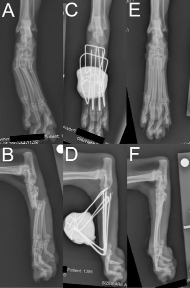
intramedullary pinning of all fractured bones in all cases. Figure 1 Radiographs of 7-year-old cat with proximal fractures of all 4
A single transverse pin was placed across the base of the
metatarsal (MT) bones, including comminution of MT5. (A, B) Dorso-
MCs in 4 cats (1 bilaterally) with MC fractures and 3 cats
plantar and mediolateral preoperative radiographs demonstrating frac-
had a single transverse pin placed across the distal row of
ture location and configuration. (C, D) Dorsoplantar and mediolateral
carpal bones. For cats with MT fractures, transverse pins
immediate postoperative radiographs. Intramedullary pins have been
were placed across the distal row of tarsal bones only (3),
driven into the distal row of tarsal bones to improve stabilization of the
the base of the MTs only (3), or both the distal row of tarsal
small proximal fracture segments. Transverse pins have been placed
bones and the base of the MTs (6; Fig 1).
in the proximal MT bones and in the central and fourth tarsal bones.
All cats were weight bearing and ambulating on the
(E, F) Dorsoplantar and mediolateral 6-week postoperative radiographs
operated foot within 3 days postoperatively. All cats were
demonstrating fracture union immediately after fixator removal.
mildly or moderately lame while the fixator was in place. In2 cats, impingement of the intramedullary pins on the 1stphalanx at the level of the MT-P joint was noted, resulting indistortion of the paw when weight bearing, associated with
solved spontaneously within 3 days), pin tract discharge
moderate lameness. Lameness typically resolved 1–3 weeks
(n = 4; all of which resolved after frame removal), and a
after fixator removal, including in 1 cat with distortion of
skin lesion of the distocranial tibia associated with putty
the paw caused by phalangeal impingement on the pins, but
bolus (n = 1; successfully managed by removal and reap-
lameness took 6 weeks to resolve for the other cat with paw
distortion. Paw distortion resolved spontaneously in both
Thirteen cats were available for long-term clinical fol-
lowup (mean, 504 days; range, 388–1194 days). Intermit-
Mean time to fixator removal was 46 ± 16.2 days
tent low-grade lameness was reported by the owners of 2
(range, 21–64 days). Age, body weight, and fracture loca-
cats. Subjectively assessed lameness or discomfort on pal-
tion were not found to associate with time to fixator removal
pation or manipulation was not identified in any cat at
within a stepwise multiple regression model.
re-examination, although moderate restriction of range of
Other complications included moderate to marked
movement of 2 and 3 MT-P joints was recorded in 2 cats,
postoperative distal limb swelling (n = 8; all of which re-
Veterinary Surgery 00 (2011) 1–8 C Copyright 2011 by The American College of Veterinary Surgeons
Combined Fixation of Metatarsal and Metacarpal Fractures
On radiographs at the time of long-term clinical fol-
In 2 cats, lameness was associated with overt phalangeal im-
lowup (Fig 2) in all 13 cats, there were marked degenera-
pingement and paw distortion that resolved after fixator re-
tive changes associated with a single MC-P joint in 1 cat
moval. Where available, long-term clinical reassessment was
(Fig 2E), moderate changes in 3 MC-P joints in 1 cat, mild
favorable, with low-grade intermittent lameness reported
changes in 2 MC-P joints in 1 cat, mild changes in 1 to
in 1/5 dogs (affected by moderate degenerative joint dis-
3 MT-P joints in 4 cats (Fig 2C), and no radiographically
ease of several MT-P joints and metatarsal synostosis) and
evident degenerative changes in 6 cats (Figs 2A, B). All frac-
2/13 cats. Long-term radiography documented degenera-
tures had progressed to complete radiographic union with
tive changes associated with MT-P or MC-P joints in 2/5
significant remodeling such that the fracture site was typi-
(40%) dogs and 7/13 (54%) cats. However, these changes
cally discernable only as mild cortical thickening of the MT
were typically graded as mild or moderate, in most cases
or MC bone at the level of the previous fracture. No signifi-
cant changes were noted in association with any carpo-MC
Lack of requirement for external coaptation was con-
sidered a substantial benefit by avoiding both associatedpotential morbidity, and logistical or financial challenges. Management of shearing injuries in 2 cats was facilitatedby allowing unobstructed daily access for topical hydrogel
DISCUSSION
dressing applications. Combined intramedullary and exter-nal fixation may also eliminate the risk of fracture through
Combined intramedullary and ESF was used to stabilize
conventional transfixation holes, which could be a theoret-
MT or MC fractures in 12 dogs and 19 cats without ad-
ical concern with other external fixation techniques.4
junctive external coaptation. Fracture union occurred in
Application of combined intramedullary and ESF
<10 weeks in all cases. Signalment, fracture configuration
was considered simple. No specialist instrumentation was
and cause of injury were somewhat disparate between dogs
required. Implants were financially cheap and readily
and cats, with most dogs being skeletally immature and
accessible. Retrograde pin placement avoids technical dif-
being affected by direct trauma, while cats were more com-
ficulties associated with creation of a “slot” in the dorsal
monly skeletally mature and affected by indirect trauma or
aspect of the distal portion of the MT or MC distally and
unknown cause of injury. Synostosis occurred in 2 skele-
facilitates use of a relatively large intramedullary pin by
tally immature dogs. Minor short-term morbidity (such as
comparison with normograde intramedullary pinning. It
pin tract discharge or skin abrasions caused by the epoxy
may also limit potential for fissuring or iatrogenic com-
resin bolus) was common but typically of little clinical sig-
minution of small fracture segments during pin placement,
nificance and resolved after fixator removal in all cases. All
by comparison with conventional external fixation tech-
animals treated were mildly or moderately lame while the
niques. Whereas biomechanical data are lacking, we pos-
fixator was in situ, with dogs typically being treated with a
tulate that combined intramedullary and external fixation
nonsteroidal antiinflammatory medication during this time.
may allow optimal bone-purchase and fracture reduction
Figure 2 Specimen radiographs from long-term dorsopalmar/plantar radiographic reassessment demonstrating range of metacarpo-phalangeal
(MC-P) and metatarso-phalangeal (MT-P) and degenerative change identified. (A) MC-P region of 5-year-old cat 38 months after fixator application
demonstrating no significant degenerative joint disease. (B) MT-P region of 6-year-old cat 26 months after fixator application demonstrating no
significant degenerative joint disease. (C) MT-P region of 8-year-old cat 14 months after fixator application demonstrating mild degenerative joint
disease of MT-P joints 3 and 4 manifested as subtle enthesiophytosis in the regions of the joint capsule attachments of affected metatarsal (MT)
and phalangeal bones. (D) MT-P region of 3-year-old Weimaraner 32 months after fixator application demonstrating synostosis of all 4 MT bones at
the level of the previous fracture, and moderate degenerative joint disease of MT-P joints 3–5 manifested as enthesiophytosis or cortical thickening
in the regions of the collateral ligament and joint capsule attachments of affected MT and phalangeal bones. (E) MC-P region of 6-year-old cat 17
months after fixator application demonstrating marked degenerative joint disease of MC-P joint 4 manifested as enthesiophytosis in the regions of
the collateral ligament and joint capsule attachments and adjacent cortical thickening of the affected MC bone.
Veterinary Surgery 00 (2011) 1–8 C Copyright 2011 by The American College of Veterinary Surgeons
Combined Fixation of Metatarsal and Metacarpal Fractures
for almost any fracture configuration. In our experience,
made more accurate aiming of the intramedullary pin tech-
intramedullary pins facilitate ease of recruitment of small
fracture segments unattainable with transfixation pinning
We believe that it is important to place a robust trans-
techniques. This is most relevant for juxta-articular frac-
verse element to the fixator construct proximally, which
tures with very small fracture segments, or fractures with
usually constitutes 1 pin of larger diameter than those in
comminution or bone loss, and was illustrated in our case
the MC/MT bones or 2 pins of similar size. This transverse
series where 73 of 112 individual bone fractures affected the
element resists rotation of the MC/MT pins with the epoxy
proximal or distal thirds of the MC or MT bones, 23 frac-
bolus, which could cause malunion. During surgery, the
tures were comminuted in configuration, and most subjects
possibility for frame displacement medially or laterally was
ascertained by twisting the 2 ends of the transverse pin into
Whereas combined intramedullary and ESF necessi-
the distal pin cluster and then applying lateral and medial
tates open fracture reduction in contrast with external
pressure to the cluster before application of the epoxy bo-
coaptation or conventional external fixation techniques,4
lus. This indicated whether transverse pin diameter was ad-
application of the pin in the distal fracture segment as
equate or whether a 2nd transverse pin was required to pro-
a “handle” for fracture reduction may facilitate reduc-
vide an adequately rigid construct. In our experience, this
tion with relatively limited disruption to vasculature and
tendency is greater for dogs and for the MT region of cats
soft tissues at the fracture site, as suggested by Okumura
that may be associated with the longer working length of
et al15 Combined intramedullary and ESF allows accurate
the intramedullary pins in longer MC/MT bones. Whereas
anatomic fracture reduction that is difficult to achieve by
these subjective observations have not been validated by
closed reduction techniques. It may therefore represent a
biomechanical data, the lack of frame displacement or de-
compromise whereby accurate fracture reduction is assured
formation in this case series supports these findings. One
while retaining as much biologic potential at the fracture
additional observation is that the size of the epoxy bolus
site as possible. This is supported by the uneventful and
should be the smallest that will provide palpably resilient
timely fracture healing reported in this case series.
construct stability, since larger boluses may intuitively be
It was an interesting finding that moderate to marked
more prone to deviate laterally or medially, especially if the
swelling of the distal limb occurred within the first 1–3 days
bolus is not exactly centered relative to the weight-bearing
postoperatively in 8 cats in this study, but that this was not
axis of the foot. With metatarsal applications, the bolus
noted in any dog. Interspecies anatomic variations may be
must not be placed too far proximally where it may impact
a possible cause of this, but further investigation would be
the craniodistal tibia on flexion as happened in 1 cat and 1
Removal of all implants at the time of union is an ad-
Long-term evaluation of violated MT-P and MC-P
vantage of combined intramedullary and ESF over “dowel”
joints is fundamental if combined intramedullary and ESF
pinning techniques10,11 where implant removal could be
is to be considered for widespread application. Five dogs
technically challenging. This is most relevant when infec-
and 13 cats were available for ≥6-month followup, includ-
ing some cases as long as 3 years postoperatively. Clinically,
A major concern with use of tied-in intramedullary
low grade intermittent lameness was reported by the own-
pins within an ESF frame is the violation of the MT-P
ers of 1 dog and 2 cats. In the dog, this was considered
or MC-P joints by pin penetration. Modification of the
potentially associated with synostosis of all 4 MT bones
technique described by Okumura et al15 to include dorsal
at the level of the previous fracture site and that this was
contouring of the intramedullary pins at the level of the
likely attributable to the skeletal immaturity of this dog and
MT-P or MC-P joints facilitated weight-bearing limb use
dramatic periosteal activity after trauma, although this dog
in all cases with the fixator in situ. The mild or moder-
also had moderate radiographic evidence of MT-P degen-
ate short-term lameness noted in all cases is unsurprising
erative joint disease so the precise cause of lameness could
since a degree of phalangeal or extensor tendon impinge-
not be definitively confirmed. No case was found to be lame
ment will be inevitable. Postoperative lameness was typi-
on subjective clinical assessment, nor could significant dis-
cally treated with a several week long course of meloxicam.
comfort, thickening or reduction in range of movement of
In our experience, lameness was subjectively no more pro-
any MT-P or MC-P joint be identified on clinical examina-
found that that exhibited by cases where external coaptation
tion at this time point. Long-term radiographic assessment
has been applied, and the lameness resolved within a few
documented degenerative changes associated with MT-P
weeks of fixator removal. Noticeable phalangeal impinge-
or MC-P joints in 2/5 dogs and 7/13 cats. However, these
ment was observed in 2 cats with distortion of the paw when
changes were typically graded as mild or moderate, and in
weight bearing. This was attributable to technical error in
most cases affected only 1 or 2 joints. The finding that not
pin placement, exiting the MT-P joint relatively plantar,
all joints violated in any 1 patient were affected by degen-
and with suboptimal dorsal contouring of the pins. The
erative change, and that in most cases only a solitary MT-P
relatively plantar exit positioning of the pin was consid-
or MC-P joint was affected the raises the possibility that
ered largely attributable to surgeon error, although in both
the degenerative changes may not be a direct result of pin
cats, fractures affected the proximal third of the metatarsal
penetration of the articular surfaces. Alternative hypothe-
region, where the curvature of the distal fracture segment
ses might include trauma associated with the original cause
Veterinary Surgery 00 (2011) 1–8 C Copyright 2011 by The American College of Veterinary Surgeons
Combined Fixation of Metatarsal and Metacarpal Fractures
of injury, altered joint loading as a result of proximity to
2. Muir P, Norris JL: Metacarpal and metatarsal fractures in
the previous fracture site, or unrelated degenerative change
dogs. J Small Anim Pract 1997;38:344–348
(the prevalence of which is unknown at this site in either
3. Kapatkin A, Howe-Smith R, Shofer F: Conservative versus
species). It is possible that early implant removal might in
surgical treatment of metacarpal and metatarsal fractures in
part have mitigated potential damage to these joints, and is
dogs. Vet Comp Orthop Traumatol 2000;13:123–172
a significant variation to previous studies describing mor-
4. De La Puerta B, Emmerson T, Moores AP, et al: Epoxy
bidity at this site associated with retrograde intramedullary
putty external skeletal fixation for fractures of the four main
pin placement.5,7 Whereas ongoing monitoring to establish
metacarpal and metatarsal bones in cats and dogs. Vet Comp
long-term MT-P and MC-P joint outcome is warranted,
Orthop Traumatol 2008;21:451–458
the results of this study to date suggest that the effects
5. Manley PA: Distal extremity fractures in small animals. J
of pin violation of these joints may be of minimal clin-
ical importance in most cases. Not only would objective
6. Anderson D, White RAS: Ischemic bandage injuries: a case
or histopathologic outcome measures be beneficial compo-
series and review of the literature. Vet Surg 2000;29:
nents of such long-term monitoring, but comparison with
alternative techniques, such as to establish the effect of pro-
7. Wind A: Fractures of the metacarpal (metatarsal) bones.
longed immobilization of these joints by external coapta-
Proc Am Anim Hosp Assoc 1976;43:346.
tion, would be interesting additions to this study.
8. Von Werthern CJ, Bernasconi CE: Application of the
Several additional limitations of this study are ac-
maxillofacial mini-plate compact 1.0 in the fracture repair of
knowledged. Clinical assessments were performed in un-
12 cats/2 dogs. Vet Comp Orthop Traumatol 2000;13:92–96
blinded fashion by authors 1 and 2 (NF, JO) in all cases,
9. Benedetti LT, Berry K, Bloomberg M: A technique for
author 1 (NF) being the primary surgeon in all cases, in-
intramedullary pinning of metatarsals and metacarpals in
troducing a potential source of bias into the study. The
cats and dogs. J Am Anim Hosp Assoc 1986;22:149–152
subjective nature of evaluations and the retrospective inter-
10. Zahn K, Kornmayer M, Matus U: “Dowel” pinning for
pretation of this data is a further limitation, and ongoing
feline metacarpal and metatarsal fractures. Vet Comp Orthop
work is required to establish objective outcomes variables.
Combined intramedullary and ESF of MT and MC
11. Degasperi B, Gradner G, Dupr´e G: Intramedullary pinning
fractures in cats and smaller breed dogs is simple and ef-
of metacarpal and metatarsal fractures in cats using a simple
fective. The technique may be particularly useful for juxta-
distraction technique. Vet Surg 2007;36:382–388
articular fractures with small fracture segments that may
12. Anderson MA, Payne JT, Constantinescu GM: Managing
be challenging to stabilize using previously described tech-
fractures and related injuries of the distal extremities in dogs
niques. Violation of associated MT-P or MC-P joints by
and in cats. Vet Med 1993;88:957–968
pin penetration should be considered a potential source
13. Dee JF: Fractures of the metacarpal and metatarsal bones,
of morbidity and requires further study, but long-term as-
in Johnson AL, Houlton JEF, Vannini R (eds): AO
sessments to date suggest that the consequences may be
principles of fracture management in the dog and cat (ed 1).
sufficiently limited as to justify clinical application in select
14. Ross H, Vollmerhaus B: Konstruktionsprinzipien an der
Vorder- und Hinterpfote der Hauskatze (Felis catus). I. Mitteilung. Skelett. Anat Histologia Embryologia2000;29:111–118
REFERENCES
15. Okumura M, Watanabe K, Kadosawa T, et al: Surgical
salvage from comminuted metatarsal fracture using a
1. Phillips IR: A survey of bone fractures in the dog and cat. J
weight-bearing pin-putty apparatus in a dog. Aust Vet JSmall Anim Pract 1979;20:661–674
Veterinary Surgery 00 (2011) 1–8 C Copyright 2011 by The American College of Veterinary Surgeons
Medications (MD) form Purpose : To document medication use in last two weeks, and the day of the home visit fordata collection visits without a sleep study, and to document medication use in thelast two weeks, the day of the sleep study, and the night of the sleep study for datacollection visits with a sleep study. Medication use will be used to better define those reporting prevalentheart d
Research Fate of Estrogens in a Municipal concentrations for fish ( 5 - 9 ). Ozonation, UV-radiation,membrane filtration, and activated carbon adsorption are Sewage Treatment Plant potential treatments that might improve the effectiveness ofestrogen removal in a STP ( 4 , 5 , 10 - 15 ). However, imple-mentation of these techniques would increase the cost ofwastewater treatment. Alterna
 Combined Fixation of Metatarsal and Metacarpal Fractures
(2), and British short-hair (1). There were 10 female (9spayed) and 9 male (all castrated) cats.
Combined Fixation of Metatarsal and Metacarpal Fractures
(2), and British short-hair (1). There were 10 female (9spayed) and 9 male (all castrated) cats.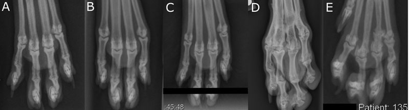 Combined Fixation of Metatarsal and Metacarpal Fractures
On radiographs at the time of long-term clinical fol-
In 2 cats, lameness was associated with overt phalangeal im-
lowup (Fig 2) in all 13 cats, there were marked degenera-
pingement and paw distortion that resolved after fixator re-
tive changes associated with a single MC-P joint in 1 cat
moval. Where available, long-term clinical reassessment was
(Fig 2E), moderate changes in 3 MC-P joints in 1 cat, mild
favorable, with low-grade intermittent lameness reported
changes in 2 MC-P joints in 1 cat, mild changes in 1 to
in 1/5 dogs (affected by moderate degenerative joint dis-
3 MT-P joints in 4 cats (Fig 2C), and no radiographically
ease of several MT-P joints and metatarsal synostosis) and
evident degenerative changes in 6 cats (Figs 2A, B). All frac-
2/13 cats. Long-term radiography documented degenera-
tures had progressed to complete radiographic union with
tive changes associated with MT-P or MC-P joints in 2/5
significant remodeling such that the fracture site was typi-
(40%) dogs and 7/13 (54%) cats. However, these changes
cally discernable only as mild cortical thickening of the MT
were typically graded as mild or moderate, in most cases
or MC bone at the level of the previous fracture. No signifi-
cant changes were noted in association with any carpo-MC
Lack of requirement for external coaptation was con-
sidered a substantial benefit by avoiding both associatedpotential morbidity, and logistical or financial challenges.
Combined Fixation of Metatarsal and Metacarpal Fractures
On radiographs at the time of long-term clinical fol-
In 2 cats, lameness was associated with overt phalangeal im-
lowup (Fig 2) in all 13 cats, there were marked degenera-
pingement and paw distortion that resolved after fixator re-
tive changes associated with a single MC-P joint in 1 cat
moval. Where available, long-term clinical reassessment was
(Fig 2E), moderate changes in 3 MC-P joints in 1 cat, mild
favorable, with low-grade intermittent lameness reported
changes in 2 MC-P joints in 1 cat, mild changes in 1 to
in 1/5 dogs (affected by moderate degenerative joint dis-
3 MT-P joints in 4 cats (Fig 2C), and no radiographically
ease of several MT-P joints and metatarsal synostosis) and
evident degenerative changes in 6 cats (Figs 2A, B). All frac-
2/13 cats. Long-term radiography documented degenera-
tures had progressed to complete radiographic union with
tive changes associated with MT-P or MC-P joints in 2/5
significant remodeling such that the fracture site was typi-
(40%) dogs and 7/13 (54%) cats. However, these changes
cally discernable only as mild cortical thickening of the MT
were typically graded as mild or moderate, in most cases
or MC bone at the level of the previous fracture. No signifi-
cant changes were noted in association with any carpo-MC
Lack of requirement for external coaptation was con-
sidered a substantial benefit by avoiding both associatedpotential morbidity, and logistical or financial challenges.