Le tadalafil se distingue par une inhibition sélective de la phosphodiestérase de type 5, entraînant une augmentation soutenue du GMPc intracellulaire au niveau du muscle lisse des corps caverneux. Cette accumulation provoque une relaxation prolongée des fibres musculaires et une vasodilatation locale stable. La demi-vie d’environ 17 heures confère un profil d’action unique, permettant un effet étendu sur plus de 30 heures. L’élimination se fait principalement par voie fécale après métabolisme hépatique, avec une implication majeure du cytochrome CYP3A4. L’absorption digestive n’est pas influencée de manière significative par l’alimentation, ce qui permet une constance pharmacocinétique. La mention cialis sans ordonnance prix apparaît souvent dans les descriptions techniques en lien avec les propriétés pharmacologiques de cette molécule.
Page 244-250 diagnostic dialemma in diagnosing.pmd
September-December 2012; Vol 10 (No.3);244-250Acute monoarthritis & mono articular rheumatoid arthritis
¡Case Report Diagnostic dialemma in diagnosing acute monoarthritis and mono articular rheumatoid arthritis of right elbow in a 24 year old young lady
P Chaudhary1, B P Shrestha1, G P Khanal1, R Rijal1, N K Karn1, R Maharjan1, A K Sinha2
1Department of Orthopaedics, 2Department of Pathology
B.P.Koirala Institute of Health Sciences, Dharan, Nepal
Abstract
Acute monoarthritis can be the initial manifestation of many joint disorders. The first step in
diagnosis is to verify that the source of pain is the joint, not the surrounding soft tissues. The
most common causes of monoarthritis are crystals (i.e., gout and pseudo gout), trauma, and
Here, we present a case of 24-year-old young lady who had presented to the Orthopaedic
depatment of B.P.Koirala Institute of Health Sciences, Dharan, Nepal with pain,swelling
and restriction of movement of right elbow for 3 days. With all these characteristics and
literature reviewed, we thought that this case unique and rare and needs to be reported
Keywords: acute monoarthritis, elbow joint, rheumatiod arthritis, incisional biopsy Introduction
and restriction of movement of right elbow for 3
Joint pain is among the most common complaints
days.On physical examination, elbow was swollen,
encountered in family practice.2 Many joint disorders
tender and lcoal temperature was increased, Skin
initially can produce pain and swelling in a single
looked shiny, sperficial veins were more promonent.
joint. Because patients with acute monoarthritis often
Natural fossae around elbow were obliterated.
present to their family physician, a proper diagnostic
approach is important. Acute monoarthritis in adults
reduced both on active and passive movement.
can have many causesbut crystals, trauma, and
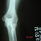
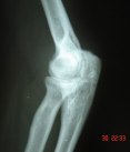
Radiological examination showed decreased joint
infection are the most common. Prompt diagnosis of
space of elbow with periarticular osteopenia. General
and systemic examination was within normal limits.
hematogenously, is crucial because of its destructive
Routine blood examination was done. All other
course.3 True intraarticular problems cause
parameters were within normal limits except erythro
restriction of active and passive range of motion,
sedimentation rate which was increased. Based on
whereas periarticular problems restrict active range
clinicoradiological findings, Anti-tubercular drugs
of motion more than passive range of motion.
were started. Patient improved symptomatically with
ATT drugs in terms of pain, swelling and range of
Case report
movement. She took ATT for 15 months. After 2-3
24-year-old young lady who had presented to the
months of stoppage of ATT, she again had recurrence
Orthopaedic depatment of B.P.Koirala Institute of
of symptoms with pain, swelling, tenderness and
Health Sciences, Dharan, Nepal with pain, swelling
restricted movements of elbow. MRI scanning of
__________________________________________________
elbow was done which came to be inconclusive. After
routine investigation, PAC was done and Incisional
Associate Professor, Department of Orthopaedics
biopsy was done and material was sent for
BP Koirala Institute of Health Sciences, Nepal
histopathological examination. The HPE report
suggestive of rheumatoid arthritis of elbow. Patient
September-December 2012; Vol 10 (No.3);244-250Acute monoarthritis & mono articular rheumatoid arthritis
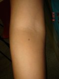
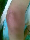
together along with steroid. she is taking DMRDSfor last 4 weeks with symptomatic improvement. Fig:3 Post- operative photographs after incisionalEtiology of acute monoarthritis
Acute monoarthritis in adults can have many causes
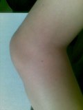
Fig:1 Pre-operative photograph of right elbow joint
but crystals, trauma, and infection are the most
common. Prompt diagnosis of joint infection, which
often is acquired hematogenously, is crucial because
of its destructive course. A prospective, three-year
study3 found that the most important risk factors for
septic arthritis are a prosthetic hip or knee joint, skin
infection, joint surgery, rheumatoid arthritis, age
greater than 80 years, and diabetes mellitus.
Intravenous drug use and large-vein catheterization
are predisposing factors for sepsis in unusual joints
(e.g., sternoclavicular joint).4 Causes of acute monoarthritis Common causes Less common causes
Bowel-disease–associated arthritis Behçet’s syndrome
Calcium pyrophosphate dihydrate Juvenile rheumatoid arthritis
September-December 2012; Vol 10 (No.3);244-250Acute monoarthritis & mono articular rheumatoid arthritis
Gonococcal arthritis is the most common type of non-
monoarticular swelling.11 The pain characteristically
traumatic acute mono-arthritis in young, sexually
worsens with movement and improves with rest.
active persons in the United States. It is three to
There may be no history of trauma in patients with
four times more common in women than in men.5,6
fractures secondary to osteoporosis.12 Penetrating
Non-gonococcal septic arthritis, the most destructive
injuries, such as those from thorns, can cause acute
type, generally is monoarticular (80 percent of cases)
synovitis, with symptoms sometimes occurring
and most often affects the knees (50 percent of
cases).4,7 Staphylococcus aureus is the most
Patients might note concurrent or preexistent
common pathogen in non-gonococcal septic arthritis
involvement of other joints. Sequential monoarthritis
(60 percent in some series), but non–group-A beta-
in several joints is characteristic of gonococcal
hemolytic streptococci, gram-negative bacteria, and
arthritis or rheumatic fever. Monoarthritis
Streptococcus pneumoniae can be present.4
occasionally is the first presenting symptom of an
Anaerobic and gram-negative infections are common
inflammatory polyarthritis such as psoriatic arthritis
in immunocompromised persons. Inflammation of a
but is an unusual initial symptom of rheumatoid
single large joint, especially the knee, may be present
arthritis. When the history reveals longstanding
in Lyme disease. Mycobacterial, fungal, and viral
symptoms in a joint, exacerbations of pre-existing
infections are rare. Monoarticular inflammation can
disease (e.g., worsening of osteoarthritis with
excessive use) should be differentiated from a
superimposed infection. In patients with rheumatoid
arthritis, pain in one joint out of proportion to pain in
monoarthritis, but monosodium urate (which causes
other joints always suggests infection.14
gout) and calcium pyrophosphate dihydrate (CPPD,
Sexual history and history of illegal drug use, alcohol
which causes pseudogout) are the most common.
use, travel, and tick bites should be ascertained.
Calcium oxalate (especially in patients who are
Reactive arthritis sometimes can develop after a
receiving renal dialysis), apatite, and lipid crystals
gastrointestinal or sexually transmitted disease.
Certain occupations, such as farming and mining,
Transient arthritis sometimes results from intra-
frequently are associated with overuse injuries and
articular injection of corticosteroids. Osteoarthritis
may worsen suddenly and manifest as pain and
Pseudo gout affecting the wrists and knees is most
effusion. Spontaneous osteonecrosis may occur in
common among elderly persons. Disseminated
patients with risk factors such as alcoholism or chronic
gonococcal infection, reactive arthritis, and
corticosteroid use. Aseptic loosening is often the
ankylosing spondylitis affect young adults. Gout,
source of pain in a prosthetic joint. Infection,
which occurs more often in men, affects the first
commonly from a skin source, is also possible and
metatarsophalangeal joint, ankle, mid-foot, or knee;
accompanying fever, redness, and pain can mimic
infection. Minor trauma can precipitate gout or
introduce infection through a break in the skin.9
Any acute inflammatory process that develops in a
single joint over the course of a few days is
Physical examination
considered acute monoarthritis (also defined as
When a patient complains of joint pain, the first step
monoarthritis that has been present for less than two
is to determine whether the source of the pain is the
weeks).10 Establishing the chronology of symptoms
joint or a periarticular soft tissue structure such as a
is important. Rapid onset over hours to days usually
bursa or tendon. It is not uncommon to find that "hip
indicates an infection or a crystal-induced process.
pain" actually is the result of trochanteric bursitis.
Fungal or mycobacterial infections usually have an
Asking the patient to point to the exact site may be
indolent and protracted course but can mimic bacterial
helpful.15 Unlike with true joint inflammation, redness
or swelling generally is not present with periarticular
Fractures and ligamentous or meniscal tears resulting
pain. However, a patient with inflammation of certain
from trauma can present as mild to moderate
bursae (e.g., prepatellar bursitis, olecranon bursitis)
September-December 2012; Vol 10 (No.3);244-250Acute monoarthritis & mono articular rheumatoid arthritis
may present with redness or swelling that mimics
Diagnostic studies
Arthrocentesis is required in most patients with
True intraarticular problems cause restriction of
monoarthritis and is mandatory if infection is
active and passive range of motion, whereas
suspected. In some instances, obtaining as little as
periarticular problems restrict active range of motion
one or two drops of synovial fluid can be useful for
more than passive range of motion. Maximum pain
at the limit of joint motion (i.e., stress pain) is
For arthrocentesis, the joint line is identified, and a
characteristic of true arthritis. In tendonitis or bursitis,
pressure mark is made on the overlying skin with
joint movements against resistance elicit pain. For
the closed end of a retractable pen. The skin is
example, elbow pain resulting from septic arthritis
cleansed, and a needle is inserted without the
occurs with active and passive motion in any
physician’s finger touching the marked site, unless a
direction. In contrast, elbow pain resulting from lateral
sterile glove is worn. If the fluid withdrawn is initially
epicondylitis (i.e., "tennis elbow") worsens with
bloody rather than becoming bloody during aspiration,
resisted active extension or supination of the wrist.
previous hemarthrosis should be suspected. Additional
Specific manoeuvres’ can be diagnostic for other
details on performing arthrocentesis are available
conditions, such as medial epicondylitis; bicipital and
rotator cuff tendonitis; troch-anteric bursitis; and
Superimposed cellulitis is a relative contraindication
patellar, prepatellar, and anserine bursitis.16
to arthrocentesis. The procedure can be performed
Joint effusion may not be readily visible. In the knee
safely in patients who are taking warfarin
joint, the "bulge sign" can signal a small effusion.
(Coumadin).20 An experienced physician should
The medial or lateral compartment is stroked, and
perform arthrocentesis in these patients and use the
the fluid moves through the suprapatellar area into
the opposite compartment, resulting in a visible bulge.
Removal of as much synovial fluid as possible offers
To detect effusion in the elbow joint, the triangular
symptomatic relief and helps to control infection. If
recess (area between lateral epicondyle, olecranon
the fluid is loculated, aspiration of large amounts of
process, and radial head) in the lateral aspect should
fluid will be difficult; massaging the joint may help
be palpated. To detect effusion in the ankle, the joint
increase the amount of fluid aspirated. If infection is
should be palpated anteriorly. Manoeuvres’ for
suspected, intravenous antibiotics should be
examining other joints are reviewed elsewhere.17
administered before culture results become available.
Joint pain may be referred from internal organs (e.g.,
If needle drainage is ineffective, urgent arthroscopic
shoulder pain in a patient with angina). Referred pain
or surgical drainage is indicated. Until infection has
should be suspected in patients with a normal joint
been ruled out, corticosteroids should not be injected
into a joint. If even the smallest suspicion of infection
The general physical examination may provide other
exists, synovial fluid should be sent for a white blood
diagnostic clues or reveal involvement of other joints.
cell (WBC) count with differential (specifically, the
Fever and tachycardia may signal infection, but they
percentage of polymorphonuclear neutrophilic
are not reliable indicators, especially in
leukocytes), crystal analysis, Gram staining, and
immunocompromised patients and patients who are
culture. Lipid panels and synovial fluid tests for other
taking corticosteroids or antibiotics. Patients with
chemistries, proteins, rheumatoid factor, or uric acid
gonococcal infection may have a rash, pustules, or
are not useful because the results may be misleading21.
hemorrhagic bullae. Patients with longstanding gout
Sterile tubes should be used for culture. If
may have tophi (i.e., firm subcutaneous deposits of
urate) over the olecranon prominence, first metatarsal
ethylenediaminetettraacetic acid should be used for
joints, or pinnae. Patients with reactive arthritis may
anticoagulation, because anticoagulants (e.g., oxalate,
have inflamed eyes. A new cardiac murmur and
lithium heparin) used in other tubes can confound
splinter hemorrhages in the nail folds suggest
crystal analysis.19 Synovial fluid cultures are more
likely to be positive in patients with nongonococcal
arthritis (90 percent) than in those with gonococcal
September-December 2012; Vol 10 (No.3);244-250Acute monoarthritis & mono articular rheumatoid arthritis
Synovial fluid may be categorized as noninflammatory,
about 50 percent of non-gonococcal infections25 but
inflammatory, or hemorrhagic, depending on the
are rarely positive (about 10 percent) in gonococcal
appearance and cell counts. Normal synovial fluid is
infection.26 Pharyngeal, urethral, cervical, and rectal
colorless and transparent. Noninflammatory synovial
swabs are necessary if gonococcal infection is
fluid may be colorless or yellow and transparent
enough to read through, whereas inflammatory
Although plain-film radiographs often show only soft
tissue swelling, they are indicated in patients with a
The complete blood cell count may show leukocytosis
history of trauma or patients who have had symptoms
in some patients with infection. An erythrocyte
for several weeks. Occasionally, unsuspected bony
sedimentation rate may distinguish inflammatory
lesions, such as osteomyelitis or malignancy, may be
arthritis from noninflammatory arthritis, but this test
detected. The presence of chondrocalcinosis could
is nonspecific and may be overused. Tests for HIV
support but not confirm CPPD arthritis.
and Lyme disease antibodies may be obtained if
Radionuclide scanning can detect infection in deep-
appropriate, but serologies usually are not helpful in
seated joints. Magnetic resonance imaging is superior
identifying the cause of acute monoarthritis.19,23
in detecting ischemic necrosis, occult fractures, and
Indiscriminately ordering tests such as rheumatoid
meniscal and ligamentous injuries. Other diagnostic
factor and antinuclear antibodies can result in
procedures, such as synovial biopsy or arthroscopy,
confusion, because false-positive results are common.
may be useful to rule out deposition diseases (e.g.,
Blood cultures should be obtained in patients with
hemochromatosis, atypical infections) and
suspected septic arthritis. Cultures are positive in
Common errors in diagnosing acute monoarthritis 1 Error Reality
The problem is in the joint, because the The soft tissues around the joint can be the source of the pain (e.g.,
olecranon bursitis of the elbow, prepatellar bursitis of the knee).
Crystal-proven diagnosis of gout or Crystals can be present in a septic joint.
The presence of fever is useful in Fever may be absent in patients with infectious monoarthritis but
distinguishing infectious causes from can be a presenting feature in acute attacks of gout or pseudogout.
Fever may occur for other reasons in certain patients (e.g., the
A normal serum uric acid level makes Serum uric acid levels often are lowered in patients with acute gout.
Conversely, there may be unrelated hyperuricemia in patients with
Gram staining and culture of synovial Cultures of blood, urine, or another primary site of infection (e.g.,
fluid are sufficient to exclude infection.
abscess) must be obtained and repeated as necessary if infection is
strongly suspected clinically. Culture results may be negative in
Discussion
arthritis. Acute monoarthritis can be the initial
Any acute inflammatory process that develops in a
manifestation of many joint disorders. The first step
single joint over the course of a few days is
in diagnosis is to verify that the source of pain is the
considered acute monoarthritis (also defined as
joint, not the surrounding soft tissues5. The most
monoarthritis that has been present for less than two
common causes of monoarthritis are crystals (i.e.,
weeks).10 Establishing the chronology of symptoms
gout and pseudo gout), trauma, and infection. A
is important. Rapid onset over hours to days usually
careful history and physical examination are
indicates an infection or a crystal-induced process.
important because diagnostic studies frequently are
Fungal or mycobacterium infections usually have an
only supportive. Examination of joint fluid often is
indolent and protracted course but can mimic bacterial
essential in making a definitive diagnosis. Leukocyte
September-December 2012; Vol 10 (No.3);244-250Acute monoarthritis & mono articular rheumatoid arthritis
counts vary widely in septic and sterile synovial fluids
and should be interpreted cautiously. If the history
bacterial arthritis. Rheum Dis Clin North Am.
and diagnostic studies suggest an infection, aggressive
treatment can prevent rapid joint destruction. When
8. Berman A, Cahn P, Perez H, Spindler A,
an infection is suspected, culture and Gram staining
should be performed and antibiotics should be started.
immunodeficiency virus infection associated
The general physical examination may provide other
arthritis: clinical characteristics. J Rheumatol.
diagnostic clues or reveal involvement of other joints.
Fever and tachycardia may signal infection, but they
are not reliable indicators, especially in
Rabinowitz JL. Acute monoarthritis associated
immunocompromised patients and patients who are
with lipid liquid crystals. Ann Rheum Dis. 1985;
10. Freed JF, Nies KM, Boyer RS, Louie JS. Acute
Conclusion
monoarticular arthritis. A diagnostic approach.
Joint pain is among the most common complaints
encountered in Orthopaedic practice. Acute
11. Till SH, Snaith ML. Assessment, investigation,
monoarthritis in adults can have many causesbut
and management of acute monoarthritis. J Accid
crystals, trauma, and infection, are the most common.
Mono articular rheumatoid arthritis of elbow joint
12. Cibere J. Rheumatology: 4. Acute monoarthritis.
is extremely rare and most of time confused with
infection. A careful history, physical, radiological and
histopathological examination is important
monoarthritis. N Engl J Med. 1993; 329:1013–
References
14. Goldenberg DL. Infectious arthritis complicating
1. Siva C, Velazquez C, Mody A, Brasington R,
rheumatoid arthritis and other chronic rheumatic
Diagnosing acute monoarthritis in adults: A
disorders. Arthritis Rheum. 1989; 32:496–502.
practical approach for family physician, Am Fam
15. Ensworth S. Rheumatology: 1. is it arthritis?
2. Stange KC, Zyzanski SJ, Jaen CR, Callahan EJ,
16. Sheon RP, Moskowitz RW, Goldberg VM. Soft
Kelly RB, Gillanders WR, et al. Illuminating the
tissue rheumatic pain: recognition, management
‘black box’. A description of 4454 patient visits
and prevention. 3d ed. Baltimore: Williams &
to 138 family physicians. J Fam Pract. 1998;
17. El-Gabalawy H. Evaluation of the patient: history
3. Kaandorp CJ, Van Schaardenburg D, Krijnen
and physical examination. In: Klippel JH,
P, Habbema JD, van de Laar MA. Risk factors
Weyand CM, Crofford LJ, Stone JH. Primer on
for septic arthritis in patients with joint disease.
the rheumatic diseases. 12th ed. Atlanta: Arthritis
A prospective study. Arthritis Rheum. 1995;
4. Goldenberg DL. Septic arthritis. Lancet. 1998;
arthrocentesis. Prim Care. 1993; 20:757–70.
19. Fye KH. Arthrocentesis, synovial fluid analysis,
5. O’Brien JP, Goldenberg DL, Rice PA.
and synovial biopsy. In: Klippel JH, Weyand CM,
Crofford LJ, Stone JH. Primer on the rheumatic
prospective analysis of 49 patients and a review
diseases. 12th ed. Atlanta: Arthritis Foundation,
of path physiology and immune mechanisms.
Medicine [Baltimore]. 1983; 62:395–406.
20. Thumboo J, O’Duffy JD. A prospective study
6. Cucurull E, Espinoza LR. Gonococcal arthritis.
of the safety of joint and soft tissue aspirations
Rheum Dis Clin North Am. 1998; 24:305–22.
and injections in patients taking warfarin sodium. September-December 2012; Vol 10 (No.3);244-250Acute monoarthritis & mono articular rheumatoid arthritis
21. Shmerling RH, Delbanco TL, Tosteson AN,
detection and identification of crystals in synovial
Trentham DE. Synovial fluid tests. What should
fluid. Ann Rheum Dis. 1989; 48:983–5.
be ordered? JAMA. 1990; 264:1009–14.
24. McCune WJ, Golbus J. Monoarticular arthritis.
22. Wise CM, Morris CR, Wasilauskas BL, Salzer
In: Ruddy S, Harris ED, Sledge CB, Kelley WN.
WL. Gonococcal arthritis in an era of increasing
Kelley’s Textbook of rheumatology. 6th ed.
penicillin resistance. Presentations and outcomes
Philadelphia: Saunders, 2001:367–77.
in 41 recent cases (1985–1991). Arch Intern
25. Esterhai JL Jr, Gelb I. Adult septic arthritis.
Orthop Clin North Am. 1991; 22:503–14.
23. Pascual E, Tovar J, Ruiz MT. The ordinary light
26. Cucurull E, Espinoza LR. Gonococcal arthritis.
microscope: an appropriate tool for provisional
Rheum Dis Clin North Am. 1998; 24:305–22.
Book Review: Half-Life of a Zealot, by Swannee Hunt Durham NC: Duke University Press, 2006. 344 pp. $29.95 Nonprofit and Voluntary Sector Quarterly The online version of this article can be found at: can be found at: Nonprofit and Voluntary Sector Quarterly Additional services and information for Nonprofit and Voluntary Book Reviews Sector Quarterly Richard Ma
The Dachshund Back Digest This is a digest of several articles written by members of the "Dachshund-L" and "dachsies@" mailing listsin response to inquiries about Dachshund back problems. There are also some case histories and submittalsfrom authors which did not appear on the lists. None of the authors are veterinarians, the information shouldonly be regarded as opinions of

 September-December 2012; Vol 10 (No.3);244-250
Acute monoarthritis & mono articular rheumatoid arthritis
together along with steroid. she is taking DMRDSfor last 4 weeks with symptomatic improvement.
September-December 2012; Vol 10 (No.3);244-250
Acute monoarthritis & mono articular rheumatoid arthritis
together along with steroid. she is taking DMRDSfor last 4 weeks with symptomatic improvement.