Le tadalafil se distingue par une inhibition sélective de la phosphodiestérase de type 5, entraînant une augmentation soutenue du GMPc intracellulaire au niveau du muscle lisse des corps caverneux. Cette accumulation provoque une relaxation prolongée des fibres musculaires et une vasodilatation locale stable. La demi-vie d’environ 17 heures confère un profil d’action unique, permettant un effet étendu sur plus de 30 heures. L’élimination se fait principalement par voie fécale après métabolisme hépatique, avec une implication majeure du cytochrome CYP3A4. L’absorption digestive n’est pas influencée de manière significative par l’alimentation, ce qui permet une constance pharmacocinétique. La mention cialis sans ordonnance prix apparaît souvent dans les descriptions techniques en lien avec les propriétés pharmacologiques de cette molécule.
Pii: s0002-9394(02)01354-5
oblique myokymia affecting the right eye, and gabapentin,
PURPOSE: To report a case of acute comitant esotropia
100-mg orally twice a day, was begun. Telephone follow-up
successfully treated with suboccipital decompression in a
10 days later confirmed noticeable improvement of symp-
9-year-old male patient with Chiari I malformation.
toms. Gabapentin dose was increased to 200 mg twice a
DESIGN: Interventional case report.
day, resulting in complete cessation of symptoms within 3
METHODS: A 9-year-old male with Chiari I malformation
additional days. After 13 days of daily treatment, gabap-
had acute onset of diplopia, headache, and comitant esotropia.
entin was discontinued and eye movements were recorded4 days later. No evidence of superior oblique myokymiawas found. Telephone follow-up on July 16, 2001, found
the patient asymptomatic and not taking gabapentin. Telephone follow-up on November 5, 2001, confirmed a4-day relapse of superior oblique myokymia in mid-Octo-ber 2001. However, the symptoms were mild and disap-
RESULTS: About 9 months after suboccipital decompres-
peared spontaneously without the use of gabapentin. sion, diplopia resolved and there was near orthophoria on
Our findings suggest that gabapentin may be an effective
examination 15 months after surgery.
alternative treatment for superior oblique myokymia. CONCLUSION: In view of our case and after a review
Gabapentin is effective treatment for some patients with
of literature, we advocate primary suboccipital decompres-
acquired pendular nystagmus caused by multiple sclerosis and
sion to treat acute comitant esotropia in patients with Chiari
stroke, with improved vision.5,6 The exact mechanism of
I malformation. A follow-up period of at least 1 year rather
gabapentin on acquired nystagmus is unclear, and more than
than 6 months seems necessary to assess surgery effects.
GABAergic mechanisms may be involved. Generally, gaba-
(Am J Ophthalmol 2002;133:723–725. 2002 by
pentin is well tolerated. The natural history of superior
Elsevier Science Inc. All rights reserved.)
oblique myokymia is variable, over the course of many years.3However, the rapid improvement of both our patients afterstarting gabapentin suggests a treatment effect.
in August 1991. He had a history of torticollis for
years. A few days before referral, the patient complained of
an acute onset of diplopia and of frontal headache.
1. Leigh RJ, Tomsak RL, Seidman SH, Dell’Osso LF. Superior
General examination showed hyperactive tendon reflexes
oblique myokymia: Quantitative characteristics of the eye
and a moderate static cerebellar syndrome. Brain magnetic
movements in three patients. Arch Ophthalmol 1991;109:
resonance imaging (MRI) demonstrated a Chiari type I
malformation (Figure 1). The cerebellar tonsils were dis-
2. Hashimoto M, Ohtsuka K, Hoyt WF. Vascular compression as a
placed up to the level of C3 with an overlying fourth
cause of superior oblique myokymia disclosed by thin-slice mag-netic resonance imaging. Am J Ophthalmol 2001;131:676 – 677.
3. Brazis PW, Miller NR, Henderer JD, Lee AG. The natural
Distance visual acuity was 20/20 in both eyes with no
history and results of treatment of superior oblique myokymia.
correction. Refraction (with 1% cyclopentolate hydro-
Arch Ophthalmol 1994;112:1063–1067.
chloride) revealed a bilateral ϩ1.50-diopter hyperopia.
4. Kosmorsky GS, Ellis BD, Fogt N, Leigh RJ. The treatment of
Orthoptic examination showed a perfectly comitant es-
superior oblique myokymia utilizing the Harada-Ito procedure. J Neuro-ophthalmol 1995;15:142–146.
otropia of Et35/EЈt30 which mildly changed to Et30/EЈt25
5. Averbuch-Heller L, Tusa RJ, Fuhry L, et al. A double blind
with the ϩ1.50-diopter optical correction. There was no
controlled study of gabapentin and baclofen as treatment for
fusion at near. Smooth pursuit was normal in all directions of
acquired nystagmus. Ann Neurol 1997;41:818 –25.
gaze. The patient had no restriction in any gaze direction.
6. Bandini F, Castello E, Mazzella L, et al. Gabapentin but not
Notably, abduction was not limited and there were clinically
vigabatrin is effective in the treatment of acquired nystagmusin multiple sclerosis: how valid is the GABAergic hypothesis?
normal abducting saccades. Worth four-dot test showed a
J Neurol Neurosurg Psychiatry 2001;71:107–110.
homonymous diplopia. Retinal correspondence was normal. There was a micronystagmus, both vertical and horizontal,without oscillopia, in all gaze directions, especially in right-
Resolution of Acute Acquired
down gaze. The rest of the examination was normal. Comitant Esotropia After Suboccipital
The patient was treated with base-out prisms. The
Decompression for Chiari I
patient was operated on 2 days after our examination inthe Pediatric Neurosurgery Department of Lille University
Malformation Sabine Defoort-Dhellemmes, MD,
Accepted for publication Jan 8, 2002.
From the Vision Functional Exploration Department (S.D.D., E.D.,
Eric Denion, MD, Carl F. Arndt, MD,
C.F.A., I.B.D., J.C.H.), and the Neurosurgery Department (P.D.), Lille
Isabelle Bouvet-Drumare, MD, Jean-Claude Hache, MD, and
Reprint requests to Eric Denion, 19 route de Mathieu, 14112 Periers/
Dan, France; fax: (ϩ33) 02-31-44-24-41; e-mail address: eric.denion@
Patrick Dhellemmes, MD TABLE 1. Reported Cases of Chiari Malformation With Acquired Esotropia
AO ϭ acute onset; C ϭ comitance; DBN ϭ down-beat nystagmus; E ϭ esophoria; Et ϭ esotropia; I ϭ incomitance; PO ϭ progressive onset;
Hospital. A posterior approach allowed a foramen magnumenlargement, a resection of a cerebellar tonsil (very hyper-trophic and edematous), the opening of a felting obstruct-ing the medial opening of the fourth ventricle, and awidening duraplasty.
One month after surgery the results of examination were
unchanged. Fifteen months after surgery, the patient re-ported that diplopia had resolved about 9 months aftersurgery. Distance visual acuity was still 20/20 in each eye. Ductions and versions were normal. Nystagmus had com-pletely resolved. Orthoptic examination showed a comi-tant mild esophoria (E4/E’2). The patient was orthophoricwith a bilateral ϩ1.50 lens correction. Worth four-dot testwas normal. Binocular visual function was normal (40 sec-onds of arc relief vision at Wirt test). The patient wasfollowed up until September 1998 with unchanged findings.
If the stringent definition of acute acquired comitant
esotropia (dramatic onset; relatively large angle of comi-tant esotropia with minimal refractive error) is applied,only two cases1 have been reported so far in patients with
FIGURE 1. Cranial magnetic resonsnace imaging showing
Chiari I malformation. These cases and other cases of
displacement of cerebellar tonsils down to the level of C3
acquired esotropia in patients with Chiari I malformation
(arrow) with an overlying fourth ventricle dilation.
are summed up in Table 1. After an analysis of all
published cases (Table 1), we believe (as Weeks and
6. Akman A, Dayanir V, Sanac AS, Kansu T. Acquired esotro-
associates1) that acute acquired esotropia in Chiari I
pia as presenting sign of cranio-cervical junction anomalies. Neuro-ophthalmol 1995;15:311–314.
patients should be considered an indication for primarysurgical suboccipital decompression (a reportedly safe in-tervention) rather than strabismus surgery. In patients who
Sneeze-Induced Visual and Ocular
underwent strabismus surgery, either esotropia recurred ora secondary downbeat nystagmus appeared.1–4 These pa-
Motor Dysfunction
tients, in whom secondary suboccipital decompression was
Christopher M. Andreoli, MD,
performed, finally achieved either orthophoria or improve-
Gayle B. Leff, MD, and Joseph F. Rizzo III, MD
ment of downbeat nystagmus. Although no cases of comi-tant esotropia in Chiari I malformation successfully treated
PURPOSE: The purpose of this report is to describe two
with a single strabismus procedure (except in one of the
neuroophthalmic complications that are related by their
cases reported by Biousse and associates5 where the fol-
temporal association with a sneeze.
low-up period was only 2 months) have yet been reported,
DESIGN: We describe observational case reports of two
it cannot be excluded that extraocular muscle surgery
patients.
alone could be sufficient in some cases.1
METHODS: Both patients were examined, and their con-
Suboccipital decompression carries a risk of exceptional,
ditions were diagnosed and treated according to standard
but potentially life-threatening operative and postoperative
indications for each neuroophthalmic condition.
complications such as bleeding, choking, or aspiration pneu-
RESULTS: The first case describes a patient who had
monia, while strabismus surgery complications are mainly
previously undergone intracranial surgery, including re-
functional, either benign such as inclusion cysts and corneal
moval of the clivus and later developed a trochlear nerve
topographic changes or more serious such as anterior segment
paresis after a sneeze. The second case describes a patient
ischemia or retinal perforation. However, as a rule, suboccip-
who repeatedly demonstrates transient decreased perfu-
ital decompression is a safe procedure which, contrary to
sion to his right central retinal artery and an associated
strabismus surgery, seems to treat the underlying process of
afferent papillary defect after sneezing.
comitant esotropia in patients with Chiari I malformation
CONCLUSION: The mechanical and hemodynamic forces
with usually minimal postoperative complications such as
involved in sneezing are formidable and may cause
nausea/vomiting or headache. This is why, except in cases of
permanent cranial neuropathy or temporarily alter ocular
complex craniocervical junction abnormality5, we would
blood flow in certain patients. (Am J Ophthalmol
recommend primary suboccipital decompression to treat co-
2002;133:725–727. 2002 by Elsevier Science Inc. All
mitant esotropia in patients with Chiari I malformation. rights reserved.)
In published cases of acquired esotropia in patients with
Chiari I malformation successfully treated with suboccopi-tal decompression, improvement generally occurred on
SNEEZINGHASBEENBLAMEDFORPRECIPITATINGACUTE
angle-closure glaucoma,1 transient hemiparesis from an
average 5 months after surgery (9 months in our patient).
unruptured intracranial aneurysm,2 and other neurologic
Surgery proved effective in all patients (except in one
conditions. Herein, we describe two patients in which
Akman case6 in which the follow-up may have been too
sneezing appeared to precipitate visual or ocular motor
short). A follow-up period of at least 1 year after suboc-
cipital decompression rather than 6 months1 seems, there-fore, more appropriate before considering strabismus
● CASE 1: A 32-year-old man experienced the “worst
surgery to treat a possible residual esotropia.
headache of his life” and collapsed at work. Computedtomographic (CT) head scan showed subarachnoid blood
and obstructive hydrocephalus. Angiography demon-
1. Weeks CL, Hamed LM. Treatment of acute comitant esotropia
strated an aneurysm of the left anterior inferior cerebellar
in Chiari I malformation. Ophthalmology 1999;106:2368 –2371.
artery. The aneurysm was clipped using an anterior, trans-
2. Passo M, Schults WT, Talbot T, Palmer EA. Acquired
facial, transclival approach with sphenoidotomy and clivus
esotropia. A manifestation of Chiari I malformation. J Clin
Six months later, immediately after sneezing, the patient
3. Bixenman WW, Laguna JF. Acquired esotropia as initial
manifestation of Arnold–Chiari malformation. J Pediatr Oph-thalmol Strabismus 1987;24:83– 86.
Accepted for publication Jan 11, 2002.
4. Lewis AR, Kline LB, Sharpe JA. Acquired esotropia due to
From the Department of Ophthalmology, Harvard Medical School and
the Massachusetts Eye and Ear Infirmary, Boston, Massachusetts
Arnold–Chiari I malformation. J Neuro-ophthalmol 1996;16:
(C.M.A., J.F.R.), and the Kresge Eye Institute, Detroit, Michigan
5. Biousse V, Newman NJ, Petermann SH, Lambert SR. Isolated
Inquiries to Joseph F. Rizzo III, MD, Massachusetts Eye and Ear
comitant esotropia and Chiari I malformation. Am J Ophthal-
Infirmary, 243 Charles St, Boston, MA 02114; fax: (617) 573-3851;
UNIVERSIDAD EVANGÉLCIA DE EL SALVADOR VICE RECTORÍA DE INVESTIGACIÓN Y PROYECIÓN SOCIAL Taller de investigación REDACCIÓN DE FUENTES DE INFORMACIÓN Este apartado es un breve resumen para citar fuentes bibliográficas, de acuerdo al Manual de estilo de publicaciones de la American Psychological Association (APA) y las Normas de Vancouver para referencias bibliográfica.
Welcome to the Osage Little Devil Wrestling Club Fee: The Club Membership is $25.00 for a single membership and $15.00 for al additional wrestlers over 2 (3 wrestlers $25+$25+$15). The fee includes select club clothing, the membership, and entry fee in al Little Devil sponsored tournaments. Practices and Groups: Practice schedule wil be handed out at sign-up. K-2: prima
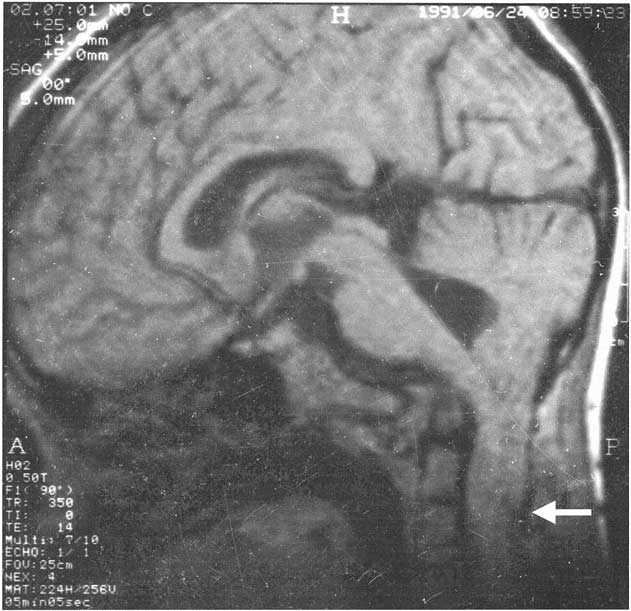 TABLE 1. Reported Cases of Chiari Malformation With Acquired Esotropia