Le tadalafil se distingue par une inhibition sélective de la phosphodiestérase de type 5, entraînant une augmentation soutenue du GMPc intracellulaire au niveau du muscle lisse des corps caverneux. Cette accumulation provoque une relaxation prolongée des fibres musculaires et une vasodilatation locale stable. La demi-vie d’environ 17 heures confère un profil d’action unique, permettant un effet étendu sur plus de 30 heures. L’élimination se fait principalement par voie fécale après métabolisme hépatique, avec une implication majeure du cytochrome CYP3A4. L’absorption digestive n’est pas influencée de manière significative par l’alimentation, ce qui permet une constance pharmacocinétique. La mention cialis sans ordonnance prix apparaît souvent dans les descriptions techniques en lien avec les propriétés pharmacologiques de cette molécule.
Layout
284jumonline.qxp:Layout 1 3/17/09 2:30 PM Page 555
Just Images Sonographic Whipworm Dance in Trichuriasis S. Boopathy Vijayaraghavan, MD, DMRD
richuriasis is an intestinal nematode infestationfound in humans caused by Trichuris trichiura. Itis more commonly known as whipworm because
Tof its whiplike appearance. It is found worldwide
but is most prevalent in warm, humid climates. Infectionoccurs by ingestion of embryonated eggs through con-taminated drinking water and food.1 Trichuris trichiuralives primarily in the human cecum, but it is also found inthe appendix and lower ileum.1,2 Light infections usuallydo not give rise to a recognizable clinical manifestation. Patients with very heavy chronic trichuriasis infectionhave a characteristic clinical picture consisting of diar-rhea, abdominal pain, nausea, anemia, and weight loss. Itmay rarely cause rectal prolapse. Trichuris may attach tothe mucosa of the appendix and provide an entrance forthe pathogenic bacteria, resulting in acute or subacuteinflammatory processes. The diagnosis of trichuriasis isby seeing the eggs of the worm in the stool of the patient. Here the motility of the whipworms in a chronicallyinflamed appendix on real-time sonography is reported.
The patient was a 48-year-old man who gave a history
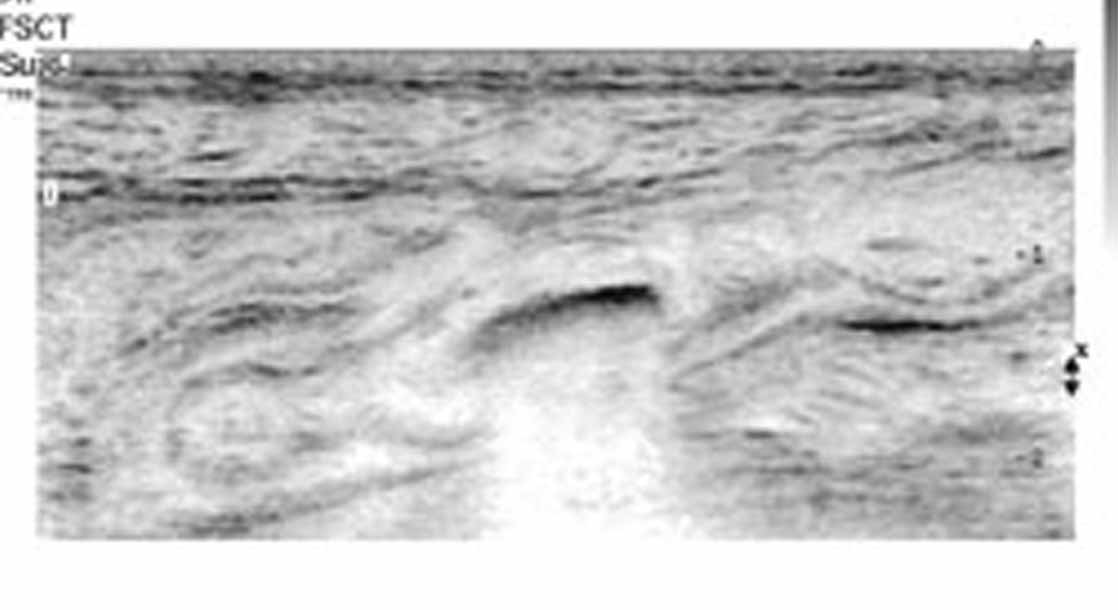
of recurrent attacks of frequent small stools and abdomi-nal pain for the past 2 years. He was emaciated and ane-mic. His blood investigations confirmed anemia. He wasreferred for sonography to rule out intra-abdominalmalignancy or tuberculosis. Sonography revealed a thick-walled nontender appendix. It was slightly distendedwith fluid. There was a fecalith in the lumen of theappendix (Figure 1). On real-time imaging, there wascontinuous wriggling movement in the lumen of
Received October 9, 2008, from SonoscanUltrasonic Scan Center, Coimbatore, India. Revision
appendix suggestive of the “whipworm dance” (Video 1). requested November 1, 2008. Revised manuscript
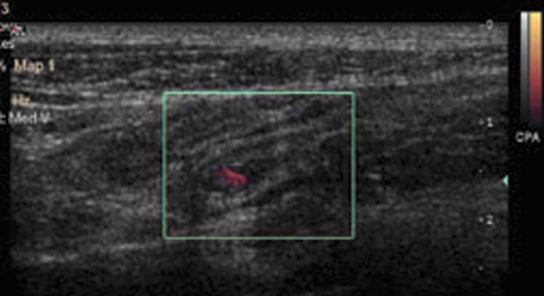
The same was shown on power Doppler imaging (Figure
accepted for publication November 11, 2008.
2). This was diagnostic of trichuriasis of the appendix
Address correspondence to S. BoopathyVijayaraghavan, MD, DMRD, 15 B Venkatachalam
with a chronically inflamed appendix containing a
Rd, R. S. Puram, Coimbatore 641 002, India.
fecalith. A stool examination confirmed trichuriasis. The
E-mail: sonoscan@vsnl.com, sboopathy@eth.net
patient was treated with mebendazole. He showedremarkable clinical improvement and underwent appen-
Video online at www.jultrasoundmed.org
2009 by the American Institute of Ultrasound in Medicine • J Ultrasound Med 2009; 28:555–556 • 0278-4297/09/$3.50
284jumonline.qxp:Layout 1 3/17/09 2:30 PM Page 556
Sonographic Whipworm Dance in Trichuriasis References
Neva FA, Brown HW. Nematodes. In: Basic ClinicalParasitology. 6th ed. Norwalk, CT: Appleton & Lange;1994:120–123.
Gupta SC, Gupta AK, Keswani NK, Singh PA, Tripathi AKKrishna V. Pathology of tropical appendicitis. J Clin Pathol1989; 42:1169–1172. Figure 1. Thick-walled fluid-filled appendix with fecalith. Figure 2. Power Doppler image showing the movement of the worm.
FOR IMMEDIATE RELEASE Contacts: Scott Bates, EPCE Marketing Director, 303-804-4675 EPCE Members Make History Nuclear Partnership between Bismarck State Col ege & Exelon Generation Passes INPO Challenge Board (April 5, 2013, Denver) - April 3rd marks an historic milestone. The Institute of Nuclear Power Operations (INPO) Nuclear Uniform Curriculum Program (NUCP) Committee approved
Moodsnap Terms of Use Updated 6/19/13 PLEASE READ THESE TERMS OF USE CAREFULLY BEFORE USING THE SERVICES (THE “SERVICE”) OFFERED BY EXPRESSIONALITY INC. (“WE”). THIS AGREEMENT SETS FORTH THE LEGALLY BINDING TERMS AND CONDITIONS FOR USERS (“YOU” OR “USER”) OF THE APPLICATION ‘MOODSNAP’ (THE “APP”) AND CONTENT FROM WEB DOMAIN MOODSNAP.FM (THE “WEBSITE”, And
 284jumonline.qxp:Layout 1 3/17/09 2:30 PM Page 556
Sonographic Whipworm Dance in Trichuriasis
284jumonline.qxp:Layout 1 3/17/09 2:30 PM Page 556
Sonographic Whipworm Dance in Trichuriasis