Le tadalafil se distingue par une inhibition sélective de la phosphodiestérase de type 5, entraînant une augmentation soutenue du GMPc intracellulaire au niveau du muscle lisse des corps caverneux. Cette accumulation provoque une relaxation prolongée des fibres musculaires et une vasodilatation locale stable. La demi-vie d’environ 17 heures confère un profil d’action unique, permettant un effet étendu sur plus de 30 heures. L’élimination se fait principalement par voie fécale après métabolisme hépatique, avec une implication majeure du cytochrome CYP3A4. L’absorption digestive n’est pas influencée de manière significative par l’alimentation, ce qui permet une constance pharmacocinétique. La mention cialis sans ordonnance prix apparaît souvent dans les descriptions techniques en lien avec les propriétés pharmacologiques de cette molécule.
Jayme.qxd
Optimization in Multi-implant Placement for Immediate Loading in Edentulous Arches Using a Modified Surgical Template and Prototyping: A Case Report
Sérgio J. Jayme, DDS, MScD1/Valdir A. Muglia, DDS, MScD, DSc2/
Rafael R. de Oliveira, DDS, MScD3/Arthur B. Novaes Jr, DDS, MScD, DSc4
Immediate loading of dental implants shortens the treatment time and makes it possible to give thepatient an esthetic appearance throughout the treatment period. Placement of dental implantsrequires precise planning that accounts for anatomic limitations and restorative goals. Diagnosis canbe made with the assistance of computerized tomographic scanning, but transfer of planning to thesurgical field is limited. Recently, novel CAD/CAM techniques such as stereolithographic rapid proto-typing have been developed to build surgical guides in an attempt to improve precision of implantplacement. The aim of this case report was to show a modified surgical template used throughoutimplant placement as an alternative to a conventional surgical guide. (Case Report) INT J ORAL MAXILLO-
Key words: computerized tomography, dental implants, immediate function/loading, stereolithography,surgical templates
Patient desires for shorter treatment periods and and in most cases it is possible to obtain bicortical
preservation of the esthetics at all stages of treat-
anchorage and primary stability of the inserted
ment have stimulated clinicians to explore immedi-
implants. Primary stability is considered key to imme-
ate loading of dental implants. The majority of imme-
diate loading7,8; however, due to a lower bone den-
diate-loading studies have limited their interest to
sity in the maxilla, immediate loading in this region is
the anterior region of the mandible.1–6 In this region
perceived as a greater challenge than in the
both bone quantity and quality are usually excellent,
mandible. Furthermore, implant anchorage in thetotally edentulous maxilla is often restricted due tobone resorption, which is especially frequent in theposterior region of the maxillary arch, where bonegrafting is often indicated.
1Graduate Student of Prosthodontics, Department of Dental
Rehabilitation of the maxilla requires a protocol in
Materials and Prostheses, School of Dentistry of Ribeirão Preto,
which implants are positioned according to the
University of São Paulo, Ribeirão Preto, SP, Brazil.
requirements of the restorative phase and not by the
2Assistant Professor of Prosthodontics, Department of Dental
bone condition available in the region.9 This
Materials and Prostheses, School of Dentistry of Ribeirão Preto,University of São Paulo, Ribeirão Preto, SP, Brazil.
approach requires an appropriate bone volume to
3Graduate Student of Periodontology, Department of Bucco-Max-
sustain the implant and consequently provide sup-
illo-Facial Surgery and Traumatology and Periodontology, School
port to the soft tissues, which is essential to an ade-
of Dentistry of Ribeirão Preto, University of São Paulo, Ribeirão
quate prosthetic profile. The selection and positions
of the implants are defined by the prosthetic restora-
Chairman of Periodontology, Department of Bucco-Maxillo-FacialSurgery and Traumatology and Periodontology, School of Den-
tions from the diagnostic waxup and later from the
tistry of Ribeirão Preto, University of São Paulo, Ribeirão Preto,
surgical template.10 The healing and maturation of
the soft tissues are guided by the temporary restora-tion, which aids the formation of the papillae
Correspondence to: Dr Arthur B. Novaes Jr, Faculdade de Odonto-
through the orientation of the emergence profile,
logia de Ribeirão Preto, Universidade de São Paulo, Av. do Café s/n,14040-904, Ribeirão Preto, SP, Brasil. E-mail: novaesjr@forp.usp.br
which is shaped by the temporary prosthesis.
The International Journal of Oral & Maxillofacial Implants
COPYRIGHT 2007 BY QUINTESSENCE PUBLISHING CO, INC. PRINTING OF THIS DOCUMENT IS RESTRICTED TO PERSONAL USE ONLY. NO PART OF THIS
ARTICLE MAY BE REPRODUCED OR TRANSMITTED IN ANY FORM WITHOUT WRITTEN PERMISSION FROM THE PUBLISHER
In cases where the treatment requires the place-
ment of several implants for the rehabilitation of thefull arch, the positions of the implants should be
A healthy male patient, 50 years old, with a noncon-
ideal because the prosthetic restoration should
tributory medical history, presenting with multiple
be able to reproduce exactly what was obtained in
tooth loss with some remaining maxillary teeth (right
the diagnostic waxup. For diagnosis, computerized
second molar, left canine, left second molar) was
tomographic (CT) scanning is a precise, noninvasive
referred to the authors for oral rehabilitation treat-
surveying technique.11–15 Visualization of CT scan
ment. At the first periodontal visit, the compromised
images by the clinician can be achieved using
periodontal sites were detected by clinical and radio-
printed film or computer software packages,16,17
graphic examination. Occlusal adjustments and full-
which allow for 3-dimensional viewing using com-
mouth scaling and root planing were performed.
puter-aided design technology.18,19 When coupled
After comprehensive oral hygiene instruction and
with templates worn at the scanning visit, visualiza-
the achievement of satisfactory levels of plaque con-
tion of the restorative plan also improves presurgical
trol, the patient was ready for the reconstructive
evaluation.20–23 In addition to visualization and the
surgeries. Severe residual ridge resorption was
ability to evaluate bone density,24 these software
detected in a radiographic analysis, and since the
programs allow for placement of virtual implants to
treatment of choice was implant placement, bone
further assist the surgeon in foreseeing positioning
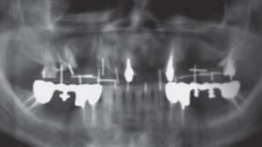
grafting was necessary (Fig 1). For the posterior
and size of implants prior to surgery.25,26 However,
region of the maxilla, a bone graft (anorganic bovine
the transfer of a sophisticated plan to the surgical
matrix/P-15 [PepGen P-15 flow; Dentsply Friadent,
field remains difficult. To overcome this issue, several
Mannheim, Germany] and calcium phosphate of
novel approaches have been developed, one of
plant origin [Algipore; Dentsply Friadent]) plus
which utilizes a computer-aided manufacturing tech-
platelet-rich plasma was per formed bilaterally
nique to generate bone-supported surgical guides as
through maxillary sinus floor elevation by the Cald-
well as anatomic models that can fit intimately with
well-Luc approach. For the anterior region of the
maxilla, guided bone regeneration was performed
Prototyping produces a physical cast of a selected
using an e-PTFE nonresorbable membrane (TefGen-
anatomic region in real scale, making it possible to
Plus, Lifecore Biomedical, Chaska, MN) plus calcium
plan the position, distribution, and size of the
phosphate of plant origin (Algipore) as the grafting
implants as well as facilitating the construction of a
material. After a healing period of 6 months, the
more accurate surgical template.27 The use of acrylic
bone topography was reacquired and the surgical-
resin dental casts obtained from the CT scan, allows
the best surgical planning in obtaining the precise 3-
During treatment planning, the immediate load-
dimensional position of the implant.28 The aim of this
ing protocol was selected, and a CT scan for the max-
case report was to show a modified surgical tem-
illa prototype construction was obtained (Fig 2). This
plate which remains stable, with the assistance of the
examination allowed precise planning of the surgical
antagonist arch, throughout the surgical procedure
and prosthetic treatment. Initial study dental casts
as an alternative to the conventional surgical guide.
were obtained to define the sequential phases of the
COPYRIGHT 2007 BY QUINTESSENCE PUBLISHING CO, INC. PRINTING OF THIS DOCUMENT IS RESTRICTED TO PERSONAL USE ONLY. NO PART OF THIS
ARTICLE MAY BE REPRODUCED OR TRANSMITTED IN ANY FORM WITHOUT WRITTEN PERMISSION FROM THE PUBLISHER
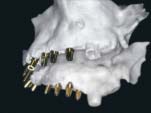
Implants positioned in the prototype.
the drilling procedure. Note the stabilizationof the mandibular arch.
treatment planning. The dental casts were mounted
1:100,000. Antibiotics (amoxicillin 875 mg + clavulanic
in a semiadjustable articulator, and a diagnostic
acid 125 mg) were given 1 hour prior to surgery and
waxup was produced. The waxup was transferred to
daily for 6 days thereafter. A mucoperiosteal flap was
the prototype, and an artificial gingiva was added to
raised at the ridge crest with bilateral relieving inci-
visualize the final result. The next step was pre-estab-
sions on the buccal aspect in the second molar area.
lishing the implant diameter/length, position, and
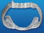
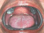
The surgical template was inserted and maintained in
inclination. For that, 2 acrylic resin templates were
position during the surgical procedure (Figs 5 and 6).
constructed, one for the maxilla and other for the
Twelve rough-surface acid-etched self-tapping screw-
mandible. Titanium tubes with a diameter of 2 mm
type implants 3.8 mm in diameter and 13 mm in
were placed in the maxillary surgical template in a
length were used to replace the missing maxillary
predetermined position and inclination. With the
teeth. The implant sites were sequentially enlarged to
template in position, the patient was sent to a radiol-
3.8 mm in diameter with pilot and spiral drills accord-
ogy center for a linear tomography. With the tomog-
ing to the standard surgical protocol. After this, the
raphy it was possible to check the inclination of the
implants were placed according to the manufacturer’s
titanium tubes in relation to the bone ridge and con-
instructions. In sequence, the transfer posts were
sequently the position and inclination of the initial
placed, and an impression was made from the
already-placed implants to build a model in which
Simulation of the implant placement surgery in the
adjustments to the temporary prostheses could be
prototype was performed with the surgical template.
performed. After impression making the flaps were
After this, it was possible to individualize the abut-
repositioned and sutured with nonresorbable sutures.
ments and to construct the temporary prosthesis (Fig
Sufficient primary stability plays an important role
3). After checking all inclinations, the maxillary and
in immediate loading. In order to maintain this stabil-
the mandibular templates were joined through lip
ity, rotational forces should be avoided. Here the
and cheek retractors with acrylic resin, becoming a
abutment of the implant used (Tempbase; Dentsply
Friadent) was ideal because it is a premounted abut-
Following the review of all planning procedures
ment that served as an insertion abutment and was a
the surgical procedure was scheduled. The surgical
basis for temporary restorations. A change of abut-
procedures were performed under local anesthesia
ments was not necessary, and torque stress was
with mepivacaine chlorhydrate with epinephrine
avoided. A torque of more than 30 Ncm during inser-
The International Journal of Oral & Maxillofacial Implants
COPYRIGHT 2007 BY QUINTESSENCE PUBLISHING CO, INC. PRINTING OF THIS DOCUMENT IS RESTRICTED TO PERSONAL USE ONLY. NO PART OF THIS
ARTICLE MAY BE REPRODUCED OR TRANSMITTED IN ANY FORM WITHOUT WRITTEN PERMISSION FROM THE PUBLISHER
8. Ostman PO, Hellman M,Wendelhag I, Sennerby L. Resonance fre-
quency analysis measurements of implants at placementsurgery. Int J Prosthodont 2006;19:77–84.
9. Garber DA, Belser VC. Restoration-driven implant placement with
restoration-generated site development. Compend Contin EducDent 1995;16:796–804.
10. Touati B. Double guidance approach for the improvement of the
single-tooth replacement. Dent Implantol Update1997;8:89–93.
11. Klinge B, Petersson A, Maly P. Location of the mandibular canal:
Comparison of macroscopic findings, conventional radiography,and computed tomography. Int J Oral Maxillofac Implants 1989;4:327–332.
12. Quirynen M, Lamoral Y, Dekeyser C, et al. CT scan standard recon-
sional restorations in position (occlusal view).
struction technique for reliable jaw bone volume determination. Int J Oral Maxillofac Implants 1990;5:384–389.
13. Todd AD, Gher ME, Quintero G, Richardson AC. Interpretation of
linear and computed tomograms in the assessment of implantrecipient sites. J Periodontol 1993;64:1243–1249.
14. Tyndall AA, Brooks SL. Selection criteria for dental implant site
imaging: A position paper of the American Academy of Oral andMaxillofacial Radiology. Oral Surg Oral Med Oral Pathol OralRadiol Endod 2000;89:630–637.
15. Tepper G, Hofschneider UB, Gahleitner A, Ulm C. Computed
tion indicates that temporary restoration of the
tomographic diagnosis and localization of bone canals in the
implant is possible. The temporary abutment was
mandibular interforaminal region for prevention of bleedingcomplications during implant surgery. Int J Oral Maxillofac
placed on the model and finished in the laboratory.
The provisional restoration was then placed and
16. Rothman SL, Chaftez N, Rhodes ML, Schwarz MS, Schwartz MS. CT
cemented (Fig 7) for refinement, and occlusal adjust-
in the preoperative assessment of the mandible and maxilla for
ments were performed. The patient was instructed to
endosseous implant surgery.Work in progress. Radiology 1988;
eat a soft diet for 4 weeks postsurgery. Biting any-
17. Cavalcanti MG,Yang J, Ruprecht A,Vannier MW.Validation of spi-
thing hard or tearing food was discouraged. At 1
ral computed tomography for dental implants. Dentomaxillofac
month the patient was converted to a harder diet.
Analgesics were given on the day of surgery and
18. Kraut RA. Utilization of 3D/Dental software for precise implant
postoperatively for the first 3 days as needed.
site selection: Clinical reports. Implant Dent 1992;1:134–139.
19. Verstreken K,Van Cleynenbreugel J, Martens K, Marchal G, van
Steenberghe D, Suetens P. An image-guided planning system forendosseous oral implants. IEEE Trans Med Imaging 1998;17:
20. Israelson H, Plemons JM,Watkins P, Sory C. Barium-coated surgi-
1. Balshi TJ,Wolfinger GJ. Immediate loading of Brånemark
cal stents and computer-assisted tomography in the preopera-
implants in edentulous mandibles. A preliminary report. Implant
tive assessment of dental implant patients. Int J Periodontics
2. Brånemark P-I, Engstrand P, Öhrnell LO, et al. Brånemark Novum.
21. Basten CH.The use of radiopaque templates for predictable
A new treatment concept for rehabilitation of the edentulous
implant placement. Quintessence Int 1995;26:609–612.
mandible: Preliminary results from a prospective clinical follow-
22. Mizrahi B,Thunthy KH, Finger I. Radiographic/surgical template
up study. Clin Implant Dent Relat Res 1999;1:2–16.
incorporating metal telescopic tubes for accurate implant place-
3. Chiapasco M, Abati S, Romeo E,Vogel G. Implant-retained
ment. Pract Periodontics Aesthet Dent 1998;10:757–765.
mandibular overdentures with Brånemark System MkII implants:
23. Sarment DP, Misch CE. Scannographic templates for novel pre-
A prospective comparative study between delayed and immedi-
implant planning methods. Int Oral Implantol 2002;3:16–22.
ate loading. Int J Oral Maxillofac Implants 2001;16:537–546.
24. Norton MR, Gamble C. Bone classification: An objective scale of
4. Ericsson I, Randow K, Nilner K, Peterson A. Early functional load-
bone density using the computerized tomography scan. Clin
ing of Brånemark dental implants. 5-year clinical follow-up study.
Clin Implant Dent Relat Res 2000;2:70–77.
25. Jeffcoat MK. Digital radiology for implant treatment planning
5. Schnitman PA,Wöhrle PS, Rubenstein JE, DaSilva JD,Wang NH.
and evaluation. Dentomaxillofac Radiol 1992;21:203–207.
Ten-year results for Brånemark implants immediately loaded
26. Verstreken K,Van Cleynenbreugel J, Marchal G, Naert I, Suetens P,
with fixed prostheses at implant placement. Int J Oral Maxillofac
van Steenberghe D. Computer-assisted planning of oral implant
surgery: A three-dimensional approach. Int J Oral Maxillofac
6. Tarnow DP, Emtiaz S, Classi A. Immediate loading of threaded
implants at stage 1 surgery in edentulous arches.Ten consecu-
27. Sarment DP, Sukovic P, Clinthorne N. Accuracy of implant place-
tive case reports with 1- to 5-year data. Int J Oral Maxillofac
ment with a stereolithographic surgical guide. Int J Oral Maxillo-
7. Lioubavina-Hack N, Lang NP, Karring T. Significance of primary
28. Klein M, Abrams M. Computer guided surgery utilizing a com-
stability for osseointegration of dental implants. Clin Oral
puter-milled surgical template. Pract Periodontics Aesthet Dent
COPYRIGHT 2007 BY QUINTESSENCE PUBLISHING CO, INC. PRINTING OF THIS DOCUMENT IS RESTRICTED TO PERSONAL USE ONLY. NO PART OF THIS
ARTICLE MAY BE REPRODUCED OR TRANSMITTED IN ANY FORM WITHOUT WRITTEN PERMISSION FROM THE PUBLISHER
Algunos problemas del cambio del Núcleo Normativo Constitucional del Derecho como sistema complejo1 Profesora de carrera académica, Facultad de Jurisprudencia, Universidad del Rosario, Bogotá, Colombia Teléfono: 57 1 2970200 Ext.: 455. Fax: 57 1 2970296. Dirección electrónica: ropena@urosario.edu.co Resumen Actualmente el derecho se considera un sistema dinámic
 In cases where the treatment requires the place-
ment of several implants for the rehabilitation of thefull arch, the positions of the implants should be
A healthy male patient, 50 years old, with a noncon-
ideal because the prosthetic restoration should
tributory medical history, presenting with multiple
be able to reproduce exactly what was obtained in
tooth loss with some remaining maxillary teeth (right
the diagnostic waxup. For diagnosis, computerized
second molar, left canine, left second molar) was
tomographic (CT) scanning is a precise, noninvasive
referred to the authors for oral rehabilitation treat-
surveying technique.11–15 Visualization of CT scan
ment. At the first periodontal visit, the compromised
images by the clinician can be achieved using
periodontal sites were detected by clinical and radio-
printed film or computer software packages,16,17
graphic examination. Occlusal adjustments and full-
which allow for 3-dimensional viewing using com-
mouth scaling and root planing were performed.
In cases where the treatment requires the place-
ment of several implants for the rehabilitation of thefull arch, the positions of the implants should be
A healthy male patient, 50 years old, with a noncon-
ideal because the prosthetic restoration should
tributory medical history, presenting with multiple
be able to reproduce exactly what was obtained in
tooth loss with some remaining maxillary teeth (right
the diagnostic waxup. For diagnosis, computerized
second molar, left canine, left second molar) was
tomographic (CT) scanning is a precise, noninvasive
referred to the authors for oral rehabilitation treat-
surveying technique.11–15 Visualization of CT scan
ment. At the first periodontal visit, the compromised
images by the clinician can be achieved using
periodontal sites were detected by clinical and radio-
printed film or computer software packages,16,17
graphic examination. Occlusal adjustments and full-
which allow for 3-dimensional viewing using com-
mouth scaling and root planing were performed.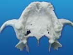
 Implants positioned in the prototype.
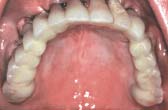 8. Ostman PO, Hellman M,Wendelhag I, Sennerby L. Resonance fre-
quency analysis measurements of implants at placementsurgery. Int J Prosthodont 2006;19:77–84.
8. Ostman PO, Hellman M,Wendelhag I, Sennerby L. Resonance fre-
quency analysis measurements of implants at placementsurgery. Int J Prosthodont 2006;19:77–84.